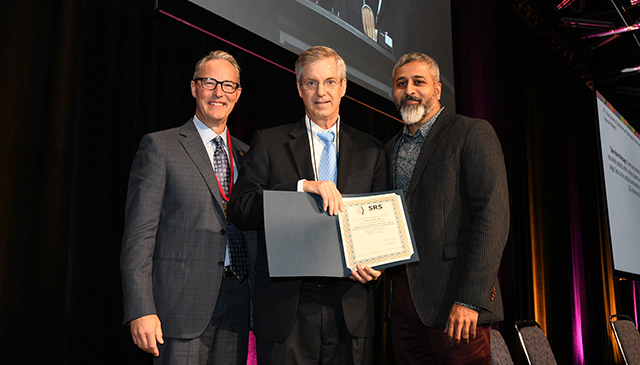
The Scoliosis Research Society (SRS) – the premier international society dedicated to the research and treatment of spinal deformities – recently honored Texas Children’s Chief of Orthopedics Dr. Brian Smith at the society’s 54th Annual Meeting, held in Montréal, Canada.
Smith received the Russell A. Hibbs Clinical Research Award, presented annually by the SRS for the meeting’s best clinical research paper. The title of the paper was “Using Proximal Humerus Ossification and Cobb Angle to Predict Progression to a Surgical Range in Adolescent Idiopathic Scoliosis Patients.”
The Hibbs Award is one of four main awards given by the SRS for basic and clinical research, and all are named for pioneers in scoliosis surgery. The SRS Program Committee selected nominees from submitted abstracts. Nominees were then invited to submit full manuscripts for review. The winners were selected based on a popular vote by meeting attendees and by committee scoring.
“I am very grateful to receive the 2019 Hibbs Award and to represent Texas Children’s Hospital in front of my peers in the SRS,” said Smith. “I would like to congratulate all my colleagues who did so much to make this happen, and I am looking forward to more research on this topic with my team at Texas Children’s.”
The paper was the result of collaborative research led by Smith and conducted with a team of experts at Yale School of Medicine. The team developed a classification system to assess skeletal maturity by analyzing proximal humerus ossification (the development of the bone of the upper arm) as seen on scoliosis patient X-rays. The study found that not only can skeletal development and maturity be reliably assessed using this system, but also that the system can be used in conjunction with other established methods to predict peak height velocity (the period when a child experiences their maximum upward growth) and the percentage of growth remaining with high accuracy. Coupled with scoliosis curve size, the system could be used as a means of predicting the risk of the patient’s potential curve progression to a surgical range during their remaining growth.
“Current systems that relied on X-ray imaging of the pelvis on scoliosis films to help determine skeletal maturation have not provided an accurate means of assessment for children with scoliosis,” Smith said. “This method has the potential to help us better define a patient’s maturity, which will help guide treatment choices and minimize cost and inconvenience of additional imaging to assess maturity.
Learn more about Texas Children’s Orthopedics Program, ranked in the top 10 nationally by U.S. News & World Report.