August 22, 2017

When Bristol Dunlap was born, she was perfectly healthy. But by the time she was three months old, she was failing to reach her milestones and began showing worrisome symptoms.

“She could not lift her head up or sit upright, her body was unusually floppy, and her left eye began turning inward even though her vision was fine,” said her mother Evonia Dunlap. “As my daughter grew older, she was slow to crawl, stand and walk, and had difficulties in chewing, swallowing and talking.”

Bristol was diagnosed with congenital hypotonia which explained her poor muscle tone throughout her body. While therapy helped her daughter sit, walk and talk, there was one thing Dunlap wanted to know: What caused Bristol to develop hypotonia?

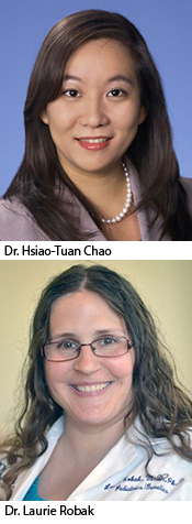
After seeing many specialists and undergoing a battery of diagnostic tests with still no answer, the family’s five-year medical odyssey ended at the Jan and Dan Duncan Neurological Research Institute (NRI) at Texas Children’s, where they were referred to neurologist Dr. Hsiao-Tuan Chao and Dr. Michael Wangler, geneticist and assistant professor of Human and Molecular Genetics at Baylor College of Medicine.

Through the NIH-funded Undiagnosed Disease Network (UDN), which brings together experts from across the country to help solve mysterious medical conditions by searching for their genetic basis, Chao and Wangler learned of a 7-year-old boy who exhibited symptoms similar to Bristol’s and also carried a point mutation in the Early B-Cell Factor 3 (EBF3) gene.

After re-examining Bristol’s exome sequencing results, they found she, too, carried the same mutation that produces a defective EBF3 protein. Since this protein is a master regulator of hundreds of other genes, even the tiniest alteration in its function could potentially cause widespread damage to the nervous system and muscles.

A referral center for undiagnosed diseases

Like the Dunlaps, many patient families are referred to Texas Children’s because of our renowned expertise and specialization in the diagnosis of diseases that are rarely seen and often unrecognized. Besides bringing together experts in genetics, pediatrics and neurology at Texas Children’s and its academic partner, Baylor College of Medicine, the UDN unites clinicians and scientists from across the country to help solve the most difficult medical cases. It is one of seven UDN sites nationwide.

More than 50 percent of UDN patients exhibit neurological symptoms. Texas Children’s chief of Neurology Dr. Gary Clark is one of the co-leaders of the UDN program at Texas Children’s and Baylor, and works closely with Texas Children’s neurologist Dr. Lisa Emrick in solving these mysterious neurological disorders.

“When a patient is referred to our UDN site, their DNA sample is submitted for sequencing,” said Emrick. “We conduct phenotyping and provide our UDN and NRI partners with the clinical patient data they need to help identify variant genes that may be responsible for a patient’s disease. Before advanced technologies like sequencing, only a small percent of these cases could be diagnosed.”

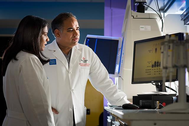
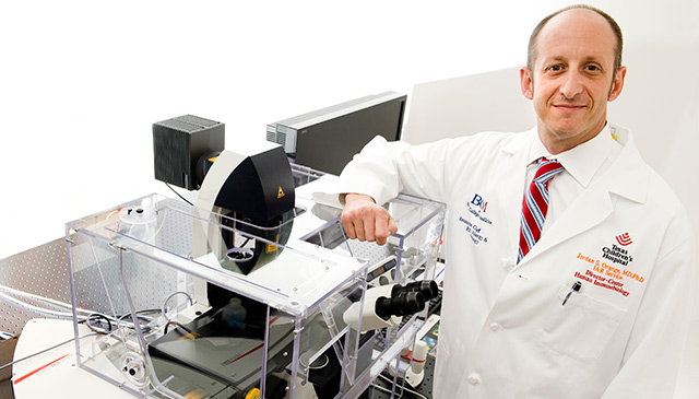
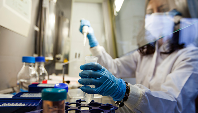
In addition to state-of-the-art medical imaging, metabolomics, and genetic testing including genome sequencing and exome sequencing, clinicians and researchers in the UDN rely on the Model Organism Screening Center (MOSC), where genes are studied in fruit flies to help diagnose patients. The center is led by Dr. Hugo Bellen, professor of Molecular and Human Genetics and Neuroscience at Baylor; Dr. Shinya Yamamoto, NRI investigator and assistant professor at Baylor; and Wangler. This dynamic team uses fruit flies, Drosophila melanogaster, to study new disease candidate genes and variants. They also closely collaborate with researchers at the University of Oregon in generating zebrafish models to study origins of disease.

In the MOSC, researchers combine bioinformatics analysis and experimentation in these ideal organism models to determine whether a specific variant identified in the genome of the patients may be responsible for the disease. “Integration of human genomics and experiments in simple model organisms such as fruit flies and zebrafish greatly facilitates disease diagnosis and mechanistic studies,” said Bellen.

“An exciting technique we developed is a way to humanize a fly gene,” said Yamamoto. “By knocking out the homologous gene in the fly and replacing it with the human gene, we can test the specific variant found in the patient to see how well it performs.”

Since fruit flies share many similar genes with humans, they have become a powerful model organism for the study of genetics. To study human disease in fruits flies, scientists mutate, or disrupt, the same gene that is known or suspected to cause the disease, and then figure out why mutations with this gene leads to disease.

Through collaboration with the UDN, Texas Children’s and Baylor continue to make significant strides in helping to accelerate the diagnoses of previously undiagnosed and rare neurological conditions.

For the Dunlap family, this provides a source of encouragement.

“While the journey to find a cure for our daughter’s illness has just begun, we are happy to finally have some answers,” Evonia said. “We are thankful for the pioneering work being done by physicians and researchers at Texas Children’s.”

June 6, 2017

Dr. Benjamin Arenkiel, a researcher at the Jan and Dan Duncan Neurological Research Institute (NRI) at Texas Children’s has received a 2017 Michael E. DeBakey, M.D., Excellence in Research Award, a prestigious honor given annually to Baylor College of Medicine faculty who have made the most significant published scientific contribution to clinical or basic biomedical research during the past three years.

“Arenkiel is an extraordinarily gifted scientist who has made many exciting and notable discoveries in the area of neuroscience,” said Dr. Huda Zoghbi, director of the NRI who nominated Arenkiel for this prestigious award. “The papers he published in the last couple of years open up new exciting areas of research into sensory maps as well as the circuits of feeding behavior.”

Since joining the Baylor faculty, Arenkiel and his team have published and/or co-authored 32 manuscripts. In addition, the Arenkiel lab now focuses on three areas of science that each have resulted in publications in high-impact journals including Developmental Cell, Neuron, Nature, and Nature Neuroscience.

Arenkiel was presented with the DeBakey Excellence in Research Award on May 15. To learn more about the scientific research that garnered Arenkiel the DeBakey award, see below:

Nature Neuroscience: Using contemporary genetic approaches, the Arenkiel lab has recently described novel roles for neuropeptide signaling in synaptic remodeling within the adult nervous system. Synapse and circuit formation and function serves as a platform for their ongoing work. Arenkiel and his lab found that, in contrast to the refinement observed for excitatory maps, inhibitory sensory maps became broader with maturation. However, like excitatory maps, inhibitory sensory maps are sensitive to experience. These data describe the development of an inhibitory sensory map as a network, highlighting the differences from previously described excitatory maps.

Developmental Cell: In this paper, Arenkiel capitalizes on the olfactory system circuits system to understand how newly born neurons integrate into an existing network in the adult brain. Using the rodent olfactory system as a model, their work strives to understand how the circuits that comprise and modulate the olfactory bulb contribute to olfaction. This research shows that local corticotropin hormone signaling onto adult-born neurons promotes and/or stabilizes chemical synapses in the olfactory bulb, revealing a neuromodulatory mechanism for continued circuit plasticity, synapse formation and integration of new neurons in the adult brain.

Nature: Most recently, Arenkiel has initiated new and creative experiments to further probe neuronal synapse and circuit formation in the mouse brain. His lab has discovered a novel mechanism by which cholinergic signaling in the basal forebrain exerts a strong influence on body weight control. His group has serendipitously uncovered novel circuits that govern convergent hypothalamic circuits that influence feeding behavior, sensory processing and stress. He has since taken a keen interest in the feeding behavior circuits. This manuscript has been positively received by the scientific community as well as the pay press and was highlighted in the Wall Street Journal.

May 30, 2017

The Clinical Research Center/Research Resources Office presented the Clinical Research Award for Second Quarter 2017 to Kathy McCarthy, research nurse, Texas Children’s Cancer and Hematology Centers.

This award was established by the Clinical Research Center in collaboration with the Research Resources Office to recognize and honor individual contributions to protecting the best interest of the research subjects and compliance with applicable rules and regulations.

McCarthy’s research activities in the CRC focus on conducting clinical trials of new agents to treat pediatric cancers. She actively participates in the screening and management of patients enrolled on Phase I and II clinical trials, as well as supportive care for these patients and their families.

May 23, 2017

More than 300 members of the Texas Children’s Hospital Department of Surgery attended the eighth annual Edmond T. Gonzales, Jr., Surgical Research Day on May 19. The event provided a forum for researchers across the department to showcase their work.

“As the research enterprise within the Department of Surgery grows, this day, where we highlight the research being conducted in the department, is more important than ever,” said Dr. Charles D. Fraser, Jr., surgeon-in-chief at Texas Children’s Hospital. “The new research discoveries coming from academic surgeons and scientists at Texas Children’s Hospital are changing the future of health care.”

This year 159 abstracts were submitted for review. Eleven of these abstracts were chosen for oral presentations on Surgical Research Day and 130 were accepted for poster presentations. This year eight students vied for the Best Presentation award and three faculty members were chosen to present their research.

The 2016 keynote speaker was Dr. Michael Longaker, the Deane P. and Louise Mitchell Professor and Vice Chair of the Department of Surgery at the Stanford University School of Medicine and Lucile Salter and Packard Children’s Hospital. He is a pediatric plastic surgeon who also directs Stanford Medical School’s Program in Regenerative Medicine and Children’s Surgical Research, and co-directs the Institute of Stem Cell Biology and Regenerative Medicine.

Laura Laux Higgins, director of special projects in the Department of Surgery at Texas Children’s Hospital, gave an ethics presentation on ethical violations in research. Higgins worked at leading legal firms as a litigator and spent several years at a top management consulting firm before joining Texas Children’s.

Awards presented at the event were the Samuel Stal Research Award for outstanding research by a resident or fellow, the Research Mentor Award, and Best Oral Presentation and Best Poster Presentation.

  • The Samuel Stal Research Award was presented to Dr. Luis De Leon, a post-doctoral research fellow in Congenital Heart Surgery. The award is named after Dr. Samuel Stal, former chief of Plastic Surgery at Texas Children’s.
  • Congenital Heart Surgeon Dr. Carlos Mery was honored with the Research Mentor Award. The award is given on an annual basis to honor a Department of Surgery faculty member who serves as a research mentor through career development, professional guidance or cultivation of research interests.
  • Best Oral Presentation award was given to Dr. James Fisher who is in the first year of his fetal surgery fellowship. He presented his work on the Development and Validation of a Fetal 3-D Surgical Simulator: Implications for Minimally Invasive In-Utero Gastroschisis Repair.
  • Best Poster award was given to researcher Andrew Lee from Anesthesiology for a poster on Use of the Baxter Faces (BARF) Scale to Measure the Severity of Nausea in Spanish Speaking Children.

Recipients of the 2017 Surgical Seed Grant Awards were announced as the grand finale of Texas Children’s Hospital Surgical Research Day. This grant program, funded by the Department of Surgery, allows surgery researchers to generate the preliminary data necessary for National Institutes of Health (NIH) grant applications and other extramural funding. Click here to find out who received a seed grant.

The Surgical Research Day Planning Committee included Dr. Jed Nuchtern (chair), Melinda Mathis (co-chair), Dr. Swathi Balaji, Shon Bower, Kathy Carberry, Dr. Jennifer Dietrich, Matthew Girotto, Laura Laux Higgins, Dr. Chester Koh, Dr. Lingkun Kong, Dr. Sandi Lam, Liz McCullum, Anissa Quiroz, Angie Rangel, Dr. Scott Rosenfeld, Stacy Staples and Veronica Victorian.

Drs. Hsiao-Tuan Chao and Laurie Robak, physician-scientists at the Jan and Dan Duncan Neurological Research Institute (NRI) at Texas Children’s, received scholarship grants from the American Academy of Neurology (AAN) to support studies on neurodevelopmental and neurodegenerative diseases.

Chao, a postdoctoral associate in the laboratory of Dr. Hugo J. Bellen was awarded the 2017 Neurology Research Training Scholarship for her proposed study titled, “Transcriptional Dysregulation of Neural Circuits in Neurodevelopmental Disorders.” Using the laboratory fruit fly and mouse, she will explore how changes in the function of master regulators of gene expression, like EBF3, can cause childhood neurologic diseases. Chao’s discoveries will provide some answers and improve the quality of life for many of these children and families.

Robak is a postdoctoral fellow in the laboratory of Dr. Joshua Shulman. She was awarded the 2017 Clinical Research Training Fellowship in Parkinson’s Disease for her proposal titled, “Elucidating Genetic Links Between Lysosomal Storage Disorders and Parkinson’s Disease.” Her study will identify lysosomal storage disorder genes as risk factors for Parkinson’s Disease, which will hopefully lead to improved diagnosis and risk assessment, and development of novel therapeutic strategies.

Twenty award winners, including Drs. Chao and Robak, were recognized at the 69th Annual Meeting of AAN, the world’s largest association of neurologists in April.

Click here for more information about their proposed research studies.

May 16, 2017

Dr. Sundeep Keswani, surgical director of Basic Research and pediatric surgeon at Texas Children’s Hospital and associate professor of surgery in the Division of Pediatric Surgery at Baylor College of Medicine, was recently awarded a $300,000 grant over a period of three years for his project “Targeting the Extracellular Matrix: an Innovative Strategy to Improve Pulmonary Hypertension in Congenital Diaphragmatic Hernia.”

The award was from the March of Dimes Foundation, which supports research consistent with its mission to improve the health of babies by preventing birth defects, premature birth and infant mortality. Less than 10 percent of applications submitted annually to the foundation are awarded.

The focus of Keswani’s awarded research is to understand the molecular mechanisms of neonatal pulmonary hypertension and develop new innovative therapies for these patients.

“The data and ideas in this application were developed here at Texas Children’s and are a direct result of the support we have received from the Department of Surgery and the hard work of our team,” Keswani said. “This work illustrates the need for surgeons to engage in basic science research to take observations from the bedside to the bench with the overall goal of improving patient outcomes.”

Keswani is a member of the pediatric surgery and fetal surgery teams at Texas Children’s and the principal investigator for the Texas Children’s Laboratory for Regenerative Tissue Repair. His NIH-funded laboratory was launched about two years ago and spans all 10 surgical divisions at Texas Children’s. Researchers in the lab study the molecular mechanisms of regenerative fetal tissue repair and are actively developing novel therapeutics to achieve postnatal regenerative wound healing.

“Conducting research is essential to provide new techniques and treatments for children’s surgery,” said Texas Children’s Surgeon-in-Chief Dr. Charles D. Fraser Jr. “Dr. Keswani’s recent award from the March of Dimes is an example of how the Department of Surgery is continuing to grow in this area, bringing in new funding and contributing significant research findings. We are committed to basic and translational research by supporting surgeon-scientists at Texas Children’s Hospital.”

Other recent awards garnered by researchers in the Regenerative Tissue Repair Lab include:

Balaji receives Wound Healing Foundation Research Grant

Dr. Swathi Balaji received the 2017 Wound Healing Foundation-FLASH Clinical Wound Healing Grant Award for her proposal titled “Pathogenesis of Cutaneous Fibrosis and Scarring.”

It is unknown why some individuals heal with robust fibrosis and scarring while others heal from similar injuries with less scarring. Balaji and her colleagues want to understand how immunoregulatory factors, particularly lymphocytes, make decisive contributions to dermal fibrosis. They propose that there are fundamental biologic differences in how fibroblasts and lymphocytes crosstalk to influence scar formation in different people. At the completion of this study, their team hopes to better understand how inflammation shapes scar formation and start working towards the development of innovative tools to promote immune regulatory responses in wounds to prevent dermal scarring as well as help other disease processes characterized by excessive fibroplasia.

The Wound Healing Foundation (WHF), through the support of the Wound Reach Foundation presented this award to Balaji at the 2017 Wound Healing Society Annual Meeting in San Diego.

Basic science research conducted by Balaji received national awards

Balaji was this year’s recipient of the ACell Young Investigator Faculty Award presented to a junior faculty member for a research abstract at the Regenerative Medicine Workshop at Hilton Head. Balaji presented a keynote lecture on her work titled “Effect of Stretch on Extracellular Matrix and Morphology of Fibroblasts in Regenerative Wound Healing.”

Tissue repair after an injury can have a spectrum of fibrosis outcomes, and fibroblasts are the major cell type that regulates the extracellular matrix and fibrosis. Even within a single tissue, fibroblasts exhibit considerable functional diversity in response to different environmental factors such as biomechanical tension and inflammation.

Balaji and her colleagues want to explain the signaling mechanisms among fibroblasts that communicate and regulate their fibrogenic phenotype. Their group is studying the role of exosomes, which are microvesicles on the order of 30-150 nm and contain functional biomolecules such as proteins, lipids, RNA, miRNA, as biomarkers and/or targeted therapeutics to regulate the functional diversity of tissue fibroblasts and their cellular cross talk.

Dr. Monica Fahrenholtz, the postdoctoral fellow on this research project, received the Wound Healing Society trainee travel award at the conference. She gave a quick presentation at this year’s annual meeting.

May 9, 2017

Dr. Jordan Orange has been selected to receive a 2017 Michael E. DeBakey, M.D., Excellence in Research Award, a prestigious honor given annually to Baylor College of Medicine faculty who have made the most significant published scientific contribution to clinical or basic biomedical research during the past three years.

Orange’s peers – Drs. Emily Mace, Lisa Forbes and Tiphanie Vogel – nominated him for the award in honor of his work as a distinguished pediatrician-scientist who has made seminal contributions to the fields of clinical immunology, basic immunology and cell biology.

Specifically, they highlighted three papers that represent the breadth, caliber and recent impact of Orange’s work as chief of the Section of Immunology, Allergy and Rheumatology and director of the Center for Human Immunobiology. These papers were published in Nature Genetics, the Journal of Clinical Investigation and the Journal of Cell Biology and are described in greater detail below.

“Dr. Orange orchestrates a vibrant and successful clinical and basic research program applying cutting-edge cell biology to questions of important clinical relevance,” Mace said. “In addition to being a pioneer in the field of NK cell deficiency and an international authority in the study and treatment of primary immunodeficiency, he is also a world’s expert in the field of highly quantitative imaging, including super-resolution and total internal reflection microscopy.”

Mace added that Orange’s scientific accomplishments are paralleled only by his success as a leader and mentor.

“His scientific vision and innovation are combined with tireless dedication to both teaching and learning,” she said. “We all are honored to count him as a colleague.”

Orange will be presented with the DeBakey, M.D., Excellence in Research Award on May 15. To learn more about the award, click here. To learn more about the scientific research that garnered Orange the DeBakey award, see below.

Nature Genetics – Through collaboration with the Baylor Hopkins Center for Mendelian Genomics, Orange has become a leader in the discovery of novel monogenic causes of primary immunodeficiency. The discovery of COPA syndrome (Watkin et al., Nature Genetics 2015) was the result of a multi-institute collaboration led by Orange. This rare, autosomal dominant autoimmune syndrome leads to arthritis and interstitial lung disease and was identified through whole exome sequencing of affected patients and their unaffected family members. This genetic discovery was validated in Orange’s laboratory by identifying the mechanism of disease through modeling of the impacted pathway. This work was also recognized with the Lee C. Howley Sr. prize for the most outstanding paper of the year by the Arthritis Foundation in 2015.

Journal of Clinical Investigation – NK cells are innate lymphocytes that eliminate infected or diseased cells. The field of primary NK cell deficiency (NKD) is one in which Orange has been a pioneer; he penned one of the first descriptions of NKD in 2003. While rare, NKD is severe and frequently fatal. To date, four monogenic causes of isolated NKD have been published, two of these from Orange’s group. Most recently, Orange led an international team that discovered biallelic mutations in the transcription factor IRF8 are a novel cause of NKD. This paper, published in the Journal of Clinical Investigation in 2017 (Mace et al.), ended a decade-long quest to solve the original published case of NKD. Again, modeling of patient mutations in the Orange laboratory using cutting-edge cell biology revealed the mechanism of impaired NK cell development in a cohort of patients from unrelated families.

Cell Biology – As NK cells lie at the heart of Orange’s research, it is not surprising that understanding NK cell function has been a cornerstone of his basic science program. He has led the field of NK cell biology by using highly quantitative microscopy and image analysis to deeply probe their function. In the January 2017 issue of the Journal of Cell Biology, Hsu et al. describe the purpose of NK cell lytic granule convergence, a mechanism by which NK cells direct their potent cytolytic machinery to prevent damage to bystander cells. This finding, which has important clinical implications for the field of immunotherapy, featured the application of novel technology to mimic an NK cell microenvironment. This work was showcased on the cover of the journal and merited an editorial from the well-known cell biologist Dr. Ira Mellman, as well as a feature on the journal’s weekly podcast.