January 10, 2017

11117clabsi640Texas Children’s neonatal intensive care unit (NICU) 2 nursing team has a big reason to celebrate – they reached 365 days and counting with zero central line-associated bloodstream infections (CLABSI), which demonstrates the value of team work and our hospital’s commitment to cultivating an environment for safe patient care.

In November 2015, NICU 2 reported three CLABSI infections. Since then, the rate of CLABSI occurrences has dropped to zero.

“Last fall, we were concerned about the number of CLABSIs we were having not only in the NICU but across the organization,” said NICU Clinical Nurse Specialist Alex Luton. “Together, with our infection control counterparts in education and vascular access, we implemented a massive educational initiative that trained more than 2,000 nurses across the organization on several key skills in central line care.”

Besides opening dialogue with the bedside nurses and care providers to identify and close any gaps observed around central line care, current hospital policies and practices were reviewed against national standards to identify areas of care that could be standardized to reduce CLABSI occurrences.

“Prior to this initiative, units had their own way of accessing and maintaining the central line,” said NICU Education Coordinator Rachel Leva. “Now, all nurses follow the same standard procedure for central line maintenance across the hospital system which has significantly helped us reduce our CLABSI rates in NICU 2.”

Creating the new role of central line resource nurses (CLRNs) has also provided an added layer of support for CLABSI prevention. To help bedside staff focus on other important patient care responsibilities, the CLRNs round on all patients with central lines during every shift.

“By providing central line care support and education, we’ve been able to address concerns early before they potentially manifest into a bloodstream infection,” said Ivy Lynn Ersan, a CLRN in NICU 2. “For instance, if a patient sweats a lot or has a lot of secretions, we may need to check on the patient more frequently per shift and change the dressing sooner than later to ward off an infection.”

Other strategies implemented in NICU 2 to reduce CLABSI include:

  • Central Line Champions Program – Specially trained to observe and audit central line care practices, these champions serve as coaches to ensure compliance with proven CLABSI prevention strategies. While all NICU staff are trained in CLABSI prevention, central line champions undergo more intensive training and must attend monthly educational sessions.
  • Hand Hygiene – NICU 2 nurses participated in a house-wide hand hygiene competition and posted signs in the unit as a visual cue to keep hand hygiene top of mind. NICU 2 leadership worked closely with Facilities to ensure ample supply of free standing hand sanitizer in the unit.
  • Weekly CLABSI meetings – Meetings are held every Tuesday to review and share information on CLABSI occurrence and identify any gaps in practice that need to be addressed. Attendees at these meetings include members from the vascular access, infection control and CLRN teams. A representative from Facilities also attends to ensure the working environment is conducive to safe patient care.

NICU 2 Assistant Clinical Director Tanya Williams says one important aspect that helped NICU 2 nurses achieve this milestone was their questioning attitude.

“Our nurses are not afraid to ask questions when something doesn’t seem right,” Williams said. “I think fostering this culture of a questioning attitude is how we were able to get this far. I am so incredibly proud of our nurses and our CLABSI partners for helping us achieve this patient safety milestone.”

December 13, 2016

When Desiree Bradley delivered her daughter, she wasn’t sure how long the little girl would survive due to an extremely rare genetic disorder called Jarcho-Levin Syndrome, which affects the spine, ribs and respiratory system. Nine years later, however, Deonc Bradley is a sassy, joyful girl who can periodically be seen bouncing down the halls of Texas Children’s Hospital, where she sees 13 different specialists for her condition.

“If Deonc was cared for anywhere else but Texas Children’s Hospital, I truly believe she would not be here,” Desiree said. “So anything I can do to help Texas Children’s be the best place it can be, I’m willing to come out here and do.”

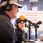
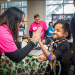
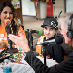
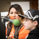
The Bradley family was one of several patient families that participated in the 2016 Texas Children’s Radiothon. Hosted by Cox Media Group Houston, the two-day event was held December 1 and 2 on the third floor of the Pavilion for Women near the Bistro Café.

Throughout the 48-hour period, people opened their hearts and their wallets donating $655,039 to the radiothon while listening to radio personalities from The Eagle (106.9 & 107.5), Country Legends (97.1) and The New 93Q (92.9) interview patients, their families and many of our clinical experts.

Texas Children’s employees gave $4,000 to the cause. Executive leadership matched those donations and added their own bringing the total amount given by Texas Children’s employees to $7,000. The Snowdrop Foundation – which was started by Kevin Kline with The Q Morning Zoo on The New 93Q, made a donation of $150,000. The foundation is dedicated to assisting patients and families at Texas Children’s Cancer Center through funding for continued research to eliminate childhood cancer and scholarships for college bound pediatric cancer patients and survivors.

“This event makes a huge difference, and it tells our story,” said Jennifer Smart, a manager in the Office of Development and the program director for the hospital’s Children’s Miracle Network Program. “We couldn’t do it without them, so of course, we’re very appreciative of that.”

View a photo gallery from the event below.

Every dollar donated to the radiothon will help Texas Children’s continue to fulfill its mission to create a healthier future for children and women throughout our global community by leading in patient care, education and research. More specifically, the money will benefit Texas Children’s Cancer Center, the Pediatric Tower expansion and the new campus Texas Children’s is building in The Woodlands.

Christi Brooks with the New 93Q and Country Legends 97.1 has participated in the radiothon since its inception and said she’s met so many families over the years who have been touched by the “miracles” that happen every day at Texas Children’s Hospital.

“Thank God we have a facility like Texas Children’s Hospital here in our back yard,” she said. “This really is truly an amazing place of miracles.”

Desiree agreed and said Texas Children’s is her and Deonc’s home away from home.

“It’s a very special place, and until you step in these walls, you don’t understand just how special this place really is.”

December 6, 2016

12716transplantinside350Seventeen-year-old Shelby Standridge came down with common colds early in her childhood, but nothing out of the ordinary. A severe nose bleed at age 9, however, landed her in the hospital and prompted questions from her parents about the cause of her unexpected illness.

Doctors in her hometown in Alabama thankfully ruled out leukemia, but did a test for cystic fibrosis, which came back positive. Cystic fibrosis is a genetic disorder that affects mostly the lungs, but also the pancreas, liver, kidneys and intestine.

At the time doctors tested Shelby for the disease, they determined her liver was already fully involved, almost to the point of end-stage liver failure. She wasn’t yet a candidate for transplant, however, so she continued battling the disease over the next five years. Throughout the past couple of years, though, Shelby’s parents and older sister noticed her lung function was declining.

“Her life had become narrow,” Shelby’s mom, Teresa Standridge, recalled.

As a result, Shelby and her family were referred to Texas Children’s, home to one of the largest pediatric transplantation programs in the nation. The team performed 86 solid organ transplants in 2015, making the program the most active pediatric transplant program in the U.S. last year.

Shelby, her mom, and older sister, Olivia, moved to Houston in October so she could be listed for a double lung and a liver transplant. A grueling wait that was expected to last six to eight months, was a mere week as Shelby underwent a 14-hour lung-liver transplant on November 8.

A multidisciplinary team of surgeons performed the procedure which is deemed so uncommon that Texas Children’s has done, including Shelby, only six of these cases since the inception of the transplant program.

“Shelby’s case is quite unusual,” said the teenager’s pulmonologist Dr. Ernestina Melicoff-Portillo. “There are only a small number of cystic fibrosis patients who have both lungs and the liver affected.”

Now, two weeks post-transplant, Shelby is feeling “ten times better” than she did and “can enjoy more in everyday life.” Her dad, Brian Standridge, noted that he hasn’t seen her grin so wide in years.

Dr. John Goss, medical director of Transplant Services, said the expertise of and the collaboration with the clinical staff and the two different surgical teams are what made Shelby’s outcome a success.

“This type of procedure only could happen at a place like Texas Children’s where our transplant program continues to earn its reputation as one of the best pediatric transplant programs in the country,” he said.

Click here to watch KHOU 11 News’ story about Shelby’s dual-organ transplant.

12716pediatricpilotawardinside900Dr. Jordan Orange, vice chair of research in the Department of Pediatrics, announced the winners of the 2016 Pediatric Pilot Awards Research Grant Program. Ten research applications were chosen by review committee members to receive grant funding in the amount of up to $50,000 for their projects.

The Pediatric Pilot Awards Research Grant Program provides initial start-up “seed funding” to support research projects. This grant program provides opportunities for new or less established researchers as well as experienced researchers who desire to expand their area of research. The grant projects are awarded based upon their scientific merit and the potential to generate the initial data necessary for a successful grant application submission to the National Institutes of Health or other external, peer-reviewed funding mechanisms.

The pilot award program is a collaborative effort between Texas Children’s Hospital and its academic partner, Baylor College of Medicine.

Congratulations to the following 2016 pilot grant awardees. View the names below to learn more about the research project being funded.

12716drsaurabhagarwal175Saurabh Agarwal, Ph.D.
Pediatrics – Hematology/Oncology
Epigenetic targeting of neuroblastoma cancer stem cells

More than half of the patients with high-risk neuroblastoma (NB) will relapse despite intensive multimodal therapy. Treatments for these patients are challenging due to disease heterogeneity, drug resistance, and toxicity. Thus, novel effective therapies are urgently required to specifically target those tumor cells which escape initial treatment and regenerate chemotherapy resistant recurrent disease.

We identified a G-CSF receptor expressing (CD114+) neuroblastoma cancer stem cell (CSC) subpopulation that is drug resistant, drives metastasis and may cause drug resistant relapse. These highly tumorigenic CSCs are distinguished by specific epigenetic alterations that lead to the expression of specific stem cell genes and maintenance of neuroblastoma CSCs.

We found that epigenetic modifiers MLL1 and JMJD3 increase the expression of G-CSF receptor gene (CSF3R) in NB CSCs by maintaining active histone modifications. Our pre-clinical studies show that blocking these epigenetic modifiers with specific small molecule inhibitors leads to neuroblastoma tumor regression and blockage of metastasis in vivo.

This pilot award will further enable us to test novel dual therapeutic approach by combining epigenetic inhibitors with standard chemotherapy for targeting both stem and non-stem neuroblastoma subpopulations. These studies will define specific epigenetic mechanisms contributing to the maintenance and tumorigenicity of NB CSCs, and pave the way for further clinical translation of our findings to block NB CSC-driven relapse and to advance a novel curative approach to neuroblastoma.
12716drwendyallenrhoades175Wendy Allen-Rhoades, M.D.
Pediatrics – Hematology/Oncology
Validation of a plasma microRNA panel as a biomarker for osteosarcoma

Osteosarcoma is the primary bone cancer in children and young adults. Currently, there are no reliable, non-invasive biological markers to detect the presence or progression of disease, assess therapy response or provide upfront prognostic insights. MicroRNAs (miRNAs) are evolutionarily conserved, stable, small non-coding RNA molecules that are key post-transcriptional regulators and are ideal candidates for circulating biomarker development due to their stability in plasma, ease of isolation and the unique expressions associated with specific disease states.

In our previous work, we analyzed more than 750 plasma miRNAs from a genetically engineered mouse model of osteosarcoma and identified a diagnostic panel of four plasma miRNAs. This diagnostic panel was able discriminate healthy from diseased animals. Subsequent analysis of 70 human patient samples corroborated these results and the diagnostic panel could discriminate healthy patients from patients with osteosarcoma. Furthermore, low plasma levels of miRNA-214 in metastatic patients at time of diagnosis were prognostic and conveyed a significantly better overall survival.

With the funding from the Pediatric Pilot Award, we will continue the necessary steps to fully validate this novel biomarker by completing validation of the diagnostic and prognostic miRNA biomarkers in 200 additional human samples. The long-term goal of this project is to test these new biomarkers in a prospective clinical trial.
12716drsaraanvari175Sara Anvari, M.D.
Pediatrics – Immunology, Allergy and Rheumatology
Defining biomarkers of successful peanut oral immunotherapy

Peanut allergy is one of the most common causes of severe and fatal allergic reactions related to food. The prevalence of peanut allergy has nearly tripled in the last 20 years and current standard of care for peanut allergy is strict avoidance of peanuts and ready access to emergency medications. While recent research has demonstrated that early introduction of peanuts, instead of avoidance, during infancy can greatly reduce the risk of a peanut allergy, this strategy is not applicable to individuals who have already developed an allergy.

For older children, teens, and adults, peanut oral immunotherapy (pOIT) is one method by which peanut allergies can be treated through step-wise introduction of peanut protein. This introduction is an effort to manage and reduce the allergic reactions in patients. However, how pOIT alters patients’ immune systems to recognize peanut protein as benign instead of “dangerous” (the nature of severe allergy) is poorly understood. Additionally, biomarkers, or testable indicators of efficacy, for pOIT success in a given patient are also still unknown at this time.

Most current research is focused on how pOIT modifies a white blood cell population, called T regulatory cells, which help control the severity of inflammation caused by immune reactions inside the human body. But T regulatory cells are just one end point of a larger set of immune reactions to pOIT. My research program will focus upstream of T regulatory cells on another population of white blood cells, called dendritic cells, which can communicate with and modify T regulatory cell, as well as several other types of white blood cells.

Specifically, the research award will help (1) identify biomarkers to predict a patient’s success (i.e. peanut tolerance) or failure (i.e. persistent peanut allergy) early in the course of pOIT, without waiting to complete three years of therapy; (2) development of targeted therapies for peanut allergic individuals aimed at altering dendritic cell populations to better modulate T regulatory populations which aid in the reduction of severe inflammatory reactions which make peanut allergies so life threatening.
12716drevelinebarbieri175Eveline Barbieri, M.D.
Pediatrics – Hematology/Oncology
Targeting MYCN-amplified neuroblastoma through RORa activation

The MYCN oncogene is a transcription factor frequently upregulated in high-risk neuroblastoma, which is profoundly involved in neuroblastoma initiation and progression. Thus, strategies antagonizing MYCN activity are a vital need in neuroblastoma therapy and the focus of this proposal.

Our laboratory has discovered that MYCN-driven neuroblastoma has an increased dependence on glutamine and lipid metabolism. Recent findings in other tumor types suggest an important link between these metabolic pathways and the circadian clock, which is disrupted in aggressive malignancies.

This led us to investigate how MYCN oncogenic signaling, circadian clock, and neuroblastoma metabolic tumor reprogramming are interrelated. Intriguingly, we have found that RORα signaling, a central component of circadian clock, is lost in MYCN-amplified neuroblastoma and this contributes to aberrant tumor proliferation.

Our specific aims will: 1) determine the metabolic programs activated by RORα in MYCN-driven tumors, and 2) determine the in vivo anti-tumor effects of RORα reactivation in pre-clinical neuroblastoma models. These studies will offer insights into critical molecular and metabolic alterations, which will provide new and more sensitive targets that could be strategically deployed with currently available therapies to treat this highly aggressive disease. Moreover, many enzymes in this pathway are amenable to small molecule inhibitors and therapeutic targeting of RORα-mediated metabolism is moving to the clinic.
12716drjennydespotovic175Jenny Despotovic, D.O.
Pediatrics – Hematology/Oncology
Genetic variants and gene expression patterns in acute and chronic immune thrombocytopenia

Immune thrombocytopenia (ITP) is an autoimmune disorder and one of the most common causes of low platelets in children. Twenty-five percent of affected children develop chronic ITP and some have significant morbidity and mortality. Currently, it is impossible to predict an individual patient’s clinical course and likelihood of spontaneous remission at the time of diagnosis.

Identification of children more likely to develop chronic ITP at diagnosis would improve treatment decisions and could also help identify important mechanisms of disease that could lead to more tailored treatment. Based on strong preliminary data produced in our laboratory, we believe that acute and chronic ITP are distinct diseases that can be distinguished at diagnosis; and specific genetic changes and gene expression differences influence the development of chronic ITP.

In our study, we are collecting DNA at enrollment on all patients with ITP, as well as RNA on patients with acute ITP at the time of diagnosis and at the time of disease resolution. For patients with chronic ITP, we are obtaining RNA at several time points. We will use the most current sequencing technologies to look for changes that may help explain differences between these two disorders with the eventual goal of identifying markers that could be used to distinguish the two disorders at diagnosis so that we could determine how to best approach each patient.
12716drjohnhollier175John Hollier, M.D.
Pediatrics – Gastroenterology
Efficacy of pre-recorded guided imagery session on pediatric gastrointestinal pain disorders managed in primary care

Up to 20 percent of school-age children and adolescents throughout the world are afflicted by recurring abdominal pain that cannot be explained by routine medical laboratory tests or procedures. These children miss more school and rank their general well-being much lower than their healthy counterparts. These disorders also may be associated with psychological distress like anxiety and depression.

One of the most effective treatments for these “functional gastrointestinal pain disorders” (FGIDs) fall under the category of cognitive behavioral therapy. However, access to this type of therapy often is not available due to lack of insurance coverage and/or scarcity of trained healthcare professionals.

Researchers have previously demonstrated the success of guided imagery, a type of cognitive behavioral therapy in treating FGIDs. Guided imagery can be delivered via using compact disc players so that patients can receive therapy at home. Our goal is to find out if audio-recorded guided imagery can be used to treat FGIDs when children are seen in the primary care setting (i.e., by their pediatrician or nurse practitioner). If so, we would be able to get treatment to these children sooner and likely decrease the need for them to be referred to a specialist (gastroenterologist). Our long term research goal is to use mobile cost effective technologies to improve the clinical care of patients with FGIDs and other pediatric diseases.
12716drandrewlandstrom175Andrew Landstrom, M.D.
Pediatrics – Cardiology
The role of junctophilin-2 in the regulation of cardiac nodal tissue

Diseases that impact the nodal tissue of the heart, such as the heart’s pacemaker, can be life-threatening. Children can suffer from these arrhythmias following surgery, through inheritance within families or for no identifiable reason. These arrhythmias can cause fainting, inability to play with the same energy as other children, or even death from collapse of the circulatory system. Despite how serious nodal disease can be, little is known about how these cells beat and how misbeats can occur. Since basic science has limited understanding of this specialized tissue, the therapies levied against nodal disease are toxic and can be ineffective.

A major reason for the lack of specialized therapies is the absence of experimental models which accurately reflect the arrhythmia. We have created an unparalleled mouse model with cardiac nodal disease that can be molecularly triggered to have arrhythmias from the nodal tissue of the heart. This mouse hosts a molecular switch which allows exposure to a pharmacological trigger to decrease the amount of a protein named junctophilin-2 (Jph2) specifically in the heart.

We have previously shown that reduction in the normal amount of Jph2 in the muscle cells of the heart causes calcium to leak into the cell. This causes a loss of contractile force and cardiac failure. We have also found that human mutations in the gene which encodes Jph2 can lead to cardiac hypertrophy as well as atrial fibrillation. All of these diseases are associated with early, and sometimes sudden, death. We have recently found that expression silencing of Jph2 specifically in the nodal tissue results in a rapid resting heart rate and an arrhythmia known as accelerated junctional rhythm. Our early studies have given strong evidence that this mouse has nodal disease that is very similar to many of the children which suffer from nodal dysrhythmias.

With support from pilot research grant, we hope to delve into the physiology of the cardiac pacemaker and to discover the molecular causes of nodal arrhythmias. We believe that the same calcium signaling that becomes perturbed in the muscle cells of the heart may be to blame for these arrhythmias. Careful interrogation of this possibility, and dissection of the molecular underpinnings of this mouse’s arrhythmias, will offer the first insights into the nodal diseases which remain unexplained and ineffectively treated.
12716drjennettemoreno175Jennette Moreno, Ph.D.
Pediatrics – Nutrition
Assessment of differences in children’s circadian rhythms during the school year and summer vacation

Consistent evidence indicates that school age children demonstrate improvements in their weight status during the school year, yet gain substantial weight during summer. These summertime increases in body mass index (BMI) increase children’s risk for becoming overweight or obese. Further, children at risk for developing chronic health conditions associated with obesity are more likely to demonstrate increases in BMI during summer. Preventing increases in children’s weight during summer may be an important opportunity to address the obesity epidemic in children.

Obesity is conventionally considered a problem of imbalance in energy intake (diet) and expenditure (physical activity/sedentary behavior). There is growing awareness of the role of sleep and circadian rhythms in the development of obesity, yet differences in children’s sleep and circadian rhythms during the school year and summer have not been examined.

Circadian rhythms are internal processes present in all living things that operate on a roughly 24 hour cycle. Behavioral rhythms such as the timing of meals and going to bed and waking up at a consistent time are some of the behaviors known to promote stable circadian rhythms. Changes in sleep and behavioral rhythms may result in disruption of circadian rhythms. Because summer vacation is associated with changes in children’s sleep and behavioral rhythms, the school year and summer vacation paradigm offer an important opportunity to expand our scientific understanding of the role of disruptions in sleep and circadian rhythms on the development of obesity in children.

Little is known about differences in children’s sleep and circadian rhythms during the school year and summer. With the current proposal, we plan to address this gap in scientific knowledge by measuring differences in children’s sleep and circadian rhythms during the school year and summer. We will assess whether differences in sleep and circadian rhythms are related to changes in children’s weight during the school year and summer. These data will may lead to novel approaches to the prevention of obesity in children.
12716drrobinparihar175Robin Parihar, M.D.
Pediatrics –Hematology/Oncology
Testing a novel non-invasive method to assess efficacy of tumor microenvironment-directed immune therapy

Some children with cancer have solid tumors, or collections of abnormally growing cells, within their organs. These collections are made up of mostly cancer cells, but also of accessory cells that help the tumor hide from the body’s immune system and grow – collectively called the tumor microenvironment. Our laboratory created a new type of immune therapy to specifically target and destroy these accessory cells found within the tumor microenvironment so that they can’t help the cancer grow.

One of the main problems for testing our immune therapy in patients with solid tumors is that we can’t detect these accessory cells without performing a biopsy procedure of the tumor inside the body. In order to detect the accessory cells in patients at many different times during their therapy, we would have to perform repeated invasive biopsy procedures, which come with additional risks and costs. There is currently no non-invasive method by which to determine the effectiveness of therapies that target the tumor microenvironment. If strategies targeting the tumor microenvironment are to be tested in humans, non-invasive methods will need to be developed to evaluate their effectiveness, thereby circumventing the need for repeated invasive biopsies.

Our project involves the creation and testing of a new type of CAT scan that can indirectly detect the accessory cells of the tumor microenvironment. If successful, our new CAT scan can be used to detect changes in the number of accessory cells in patients receiving our new immune therapy, without the need for repeated invasive, risky, or costly procedures. This new CAT scan can be used in clinical trials of other immune therapies as well and may be applied to both children and adults with cancer. The long term goal of the project is to develop a clinical imaging tool that will allow doctors to follow changes within the tumor microenvironment induced by immune therapies.
12716drsarahsartain175Sarah Sartain, M.D.
Pediatrics – Hematology/Oncology
The linkage between hemostasis-thrombosis, complement, and inflammation in the pathophysiology of thrombotic microangiopathy

The goal of our research is to improve the health of patients with thrombotic microangiopathy, a group of disorders that cause anemia, low platelets, clots in the blood vessels, and blood vessel damage of the brain, heart, and kidneys. The mechanisms of small blood vessel damage in thrombotic microangiopathy are not precisely defined.

We will investigate the means by which thrombotic microangiopathy causes blood vessel injury and organ damage. We believe that the immune system is involved in the process of vessel injury in thrombotic microangiopathy. This is based on previous work showing that components of the immune system known as the “alternative complement pathway” bind to, and become activated on, long and sticky von Willebrand factor (VWF) strings secreted from blood vessel walls. These VWF strings normally attract platelets to initiate blood clot formation. We intend to determine if activated alternative complement components on these strings contribute to blood vessel injury. We will also determine if a powerful molecule produced during inflammation (known as “tumor necrosis factor”) controls activation of the alternative complement pathway on the VWF strings, contributing to heart, brain, and kidney blood vessel injury.

Our proposed research has long-term biomedical significance because determining the mechanisms of blood vessel/organ injury in thrombotic microangiopathy will lead to the development of therapies to improve the outcomes in this disorder and may be applicable to more common types of blood vessel injury in the general population.

December 5, 2016

12516zoghbi640On Sunday, December 4, Texas Children’s and Baylor College of Medicine’s pioneering neuroscientist Dr. Huda Zoghbi was honored with the Breakthrough Prize in the field of Life Sciences during a star-studded ceremony in San Francisco’s Silicon Valley.

Awarded annually to the world’s top scientists in fundamental physics, mathematics and the life sciences, the Breakthrough Prize is considered Silicon Valley’s most significant science prize for what they cite as paradigm-shifting research. The Prize was founded by Sergey Brin (Google), Anne Wojcicki (23andMe), Mark Zuckerberg (Facebook), Priscilla Chan (Chan Zuckerberg Initiatives), Yuri Milner (DST Global) and Julia Milner.

Presented during a live broadcast on the National Geographic Channel, Zoghbi was recognized for her discoveries of the genetic causes and biochemical mechanisms of spinocerebellar ataxia, a neurodegenerative disorder affecting balance and coordination, and Rett syndrome, a genetic neurological disease characterized by the loss of motor skills, speech and other cognitive abilities affecting girls one or two years after birth. Prior to Zoghbi’s pioneering work in neuroscience, little was understood about the causes of these diseases, let alone how to potentially treat or cure them.

As one of the world’s leading neurogeneticists, Zoghbi, director of the Jan and Dan Duncan Neurological Research Institute (NRI) at Texas Children’s and Baylor, has been instrumental in other important medical breakthroughs in neurological disease research including her most recent discovery of how lowering toxic levels of tau in the brain could potentially lead to new therapies to reverse or prevent Alzheimer’s disease.

“We are tremendously proud of the transformational work Dr. Zoghbi and her team are doing at the NRI. The numerous discoveries coming from Texas Children’s and Baylor will have an immeasurable impact on so many of our families suffering from unexplainable neurological diseases,” said Texas Children’s President and CEO Mark A. Wallace. “Please join me in congratulating Dr. Zoghbi for this well-deserved and prestigious global recognition.”

Zoghbi plans to donate the majority of her $3-million Breakthrough Prize to support education and research initiatives. She wishes to recognize the institutions that impacted her career including Texas Children’s and Baylor. The gifts will help advance genetic and neuroscience research.

Zoghbi thanked her supporters with the following acceptance speech: “As a young doctor, I found it heartbreaking to watch my patients, young and old, lose their lives to neurological diseases. I turned to research for answers, and today, together with collaborators and trainees, we are charting new paths towards viable therapies. It’s thrilling to see we are beginning to understand the language of life and translate it to help mankind. I feel fortunate to have had my career nurtured by two of the finest institutions, Baylor College of Medicine and Texas Children’s Hospital and to have the support of the Howard Hughes Medical Institute. I thank my husband William and our children, Roula and Anthony, for their unwavering support, and they share this honor with me today.”

Click here to learn more about the ceremony and this award including a feature story on Zoghbi from the Houston Chronicle. FOX will air a one-hour version of the ceremony on Sunday, December 18, at 6 p.m.

In addition to this accolade, Zoghbi has earned dozens of honors and awards, including most recently the 2016 Shaw Prize in Medicine and the 2016 Jessie Stevenson Kovalenko Medal for her research.

November 29, 2016

113016surgicaloncologyinside250Texas Children’s Cancer Center is renowned for extraordinary care and outcomes and is ranked No. 2 in the nation by U.S. News & World Report. An important part of the program’s success is the multidisciplinary care children here receive – particularly in the area of surgical oncology. Many solid tumors – such as liver tumors, bone tumors, neuroblastomas and sarcomas – require complete surgical removal. Chemotherapy and radiation may be used to shrink the size of the tumor or to keep it from coming back after surgery, but surgical removal is a critical step for children with these cancers. In these cases, oncologists and surgical oncologists must work together to carefully plot the course of treatment and time the surgery just right to give young patients the very best chance at a cancer-free life.

Patient diagnosed with hepatoblastoma

In September 2015, Dr. Maria Garcia Fernandez, a pediatric infectious disease specialist, and Dr. Fernando Padilla, a family practitioner, discovered a mass in their 17-month-old baby Victoria’s abdomen. Fearing the worst, they immediately contacted the Solid Tumor Program at Texas Children’s Hospital, where Victoria was promptly evaluated and diagnosed with stage 3 hepatoblastoma.

Hepatoblastoma is a relatively rare type of childhood cancer, with approximately 200 cases diagnosed per year in the country. Usually occurring in children under the age of 5, there are often no initial symptoms other than the mass.

“Hepatoblastomas tend to present very large, because the liver is tucked under the ribs so the mass is hard to feel,” said Dr. Sanjeev Vasudevan, Victoria’s surgical oncologist specializing in liver surgery. “You have to remove the side of the liver that the tumor inhabits without damaging the normal side and still get the tumor completely out.”

The stakes for this type of surgery couldn’t be higher.

“If you attempt to remove the mass and wind up leaving some of it behind, the prognosis for the child becomes much more serious,” Vasudevan said. “Basically, if you can’t guarantee a negative-margin resection, it’s safer to skip the attempt and go straight to liver transplantation.”

Aggressive chemotherapy treatment

At the time of diagnosis, Victoria’s tumor was 6 cm in diameter and covered both sides of her liver. She had to undergo an aggressive regimen of chemotherapy to see if resection would be an option, or if transplant would be required.

“We were devastated,” Fernandez said. “We didn’t know if the chemotherapy would work, what kind of toll it would take on her, or if she’d have to have a transplant and deal with that her whole life. But, what we did know was that Texas Children’s was the best possible place for us to be. They had the numbers. They had the best track record for treating this type of cancer, whether it’s from an oncology perspective or surgery or transplant or intensive care.”

Only a handful of major centers in the country are equipped to take a case like Victoria’s. Of the 200 cases diagnosed in the U.S. annually, Texas Children’s treats approximately 10 percent of them.

“In addition to a strong cancer program, you need to have pediatric ICUs and intensivists, surgical expertise, anesthesia and pain services, all for children under the age of 5 – and enough volume to do it well and have good outcomes,” Vasudevan said.

Victoria underwent four intense cycles of chemotherapy. Each time, she was admitted back to the hospital for about a week, fighting fever, neutropenia and RSV. Knowing that four cycles was probably as much as the petite toddler could take, Victoria’s physicians were hoping to take her for surgery after one or two rounds. After the third cycle, she was placed on the transplant list briefly before scans finally showed a glimmer of hope. Victoria underwent a fourth cycle and was scanned again, and the team was delighted to find a margin of healthy tissue that made surgery possible.

“This entire team of oncologists, radiologists, pathologists, surgeons and transplant surgeons met so many times and discussed her case, all diligently trying to figure out what was best for Victoria,” Fernandez said. “It showed tremendous perseverance and dedication, and I will never forget that as long as I live.”

Surgical tumor removal

On January 6, 2016, Victoria went in for surgery, and she didn’t come out for more than nine hours. The vicinity of the tumor to the main portal vein, the primary blood supply to the liver, was close and required special attention to ensure that the tumor was completely removed.

“When operating on the liver, there is a high risk of disrupting the blood vessels and the bile ducts,” Vasudevan said. “What makes it really complicated is the fact that the liver is brown and completely opaque, and you can’t see the tumor. You rely on ultrasound guidance and external cues, the rest is up to feel and experience.”

Victoria’s procedure went smoothly. Vasudevan removed the tumor and the left lobe and was able to preserve about 60 percent of her liver.

There is a 30 percent chance of liver insufficiency post surgery, but after four or five days, the liver begins to regenerate and compensate for its loss. Victoria was stable and extubated by the next morning, and she went on to have two more cycles of chemotherapy to ensure no microscopic seeding had occurred. She has since celebrated her second birthday and returned to her normal, vibrant self.

Although Victoria is still checked regularly for signs of recurrence, overall her prognosis is excellent. She has an approximately 90 percent chance of an event-free, five-year survival.

“This is exactly why I got into this field,” Vasudevan said. “It’s an amazingly rewarding thing to do. Cancer is so devastating, in general, and to see a small 1- or 2-year-old child robbed of her whole life…that’s motivation enough for me.”

For more information about our Surgical Oncology Program, click here.

November 15, 2016

Growing up, it took awhile for Michelle Roy to warm up to other children in her neighborhood and school classrooms. A bilateral cleft lip and palate caused her to be shy, introverted and at times feel a little isolated.

“I didn’t have anyone to share my experiences and feelings with,” she said. “I didn’t have access to a network of kids in similar situations.”

Fortunately, that’s not the case for the many children Roy now works with as a physician assistant to pediatric plastic surgeon Dr. Laura Monson. Monson helped start Camp Keep Smiling in 2014, a camp for children with cleft lip and palate.

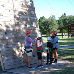
The camp recently wrapped up with a record-setting number of campers and incredible experiences for the patients and staff members alike. Camp Keep Smiling provides a safe, fun environment for patients between the ages of 10 and 16 to engage in meaningful social interaction and gain self-confidence. The camp, hosted by nonprofit Camp for All, offers activities like canoeing, fishing, archery, ropes courses, basketball and arts and crafts. Admission is free of charge for patients as it is supported directly by donations.

This year, 61 campers attended Camp Keep Smiling versus the 30 who attended in 2014. Monson leads the camp with other team members from the plastic surgery division. Physicians, nurses, OR staff and child life specialists serve as counselors who notice tremendous strides in the campers towards the end of the session. Oftentimes, campers have never met another child with cleft lip and palate and this gives them the opportunity to form friendships with those who are just like them.

“Children with cleft lip and palate often will have four or more surgeries throughout their lifetime to address not only appearance issues but speech and dental issues as well,” Monson said, adding that cleft lip and palate affects one out of every 700 births. “Camp Keep Smiling was created to help these children better understand their condition, meet people who are going through the same thing and help them deal with some of the difficult social interactions they might have growing up.”

Christy Hernandez, a registered nurse with Texas Children’s Outcomes and Impact Service, has been helping organize the camp since its inception and said it is amazing to watch the campers open up to one another over the weekend while participating in super fun activities.

“It’s life changing for many of them,” she said. “It’s a time when they can forget about their medical condition and just be kids.”

Roy participated in the camp for the first time this year as its counselor coordinator and saw firsthand what a great opportunity it is for children with cleft lips and palates to gain confidence in themselves, and meet and build relationships with people who look and often feel the same way they do.

“Camp Keep Smiling is a very valuable experience for these children to have,” she said. “I feel blessed to be a part of it and to let them know there’s a bright future ahead of them.”

Click here to watch ABC-13’s story about Camp Keep Smiling.