July 7, 2021

Seven years after performing the world’s first successful two-port fetoscopic surgery to treat spina bifida, Texas Children’s surgeons have performed the100th surgery of its kind.

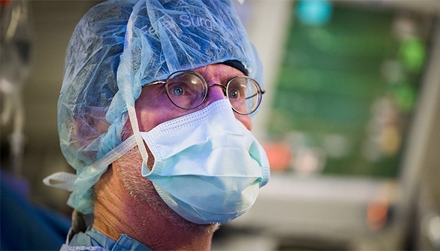
Dr. Michael Belfort, OB/GYN-in-chief at Texas Children’s Pavilion for Women, and Dr. William Whitehead, a pediatric neurosurgeon at Texas Children’s, developed the fetal surgery technique, which presents less risk to the mother and less risk of prematurity to the baby.

The condition occurs when a fetus’ neural tube fails to close during the first month of pregnancy, and can cause children to have difficulty walking, to need a shunt to drain cerebral fluid, and more.

“This milestone is very exciting and has been years in the making at Texas Children’s,” said Belfort, who is also professor and chair of the Department of Obstetrics and Gynecology at Baylor College of Medicine.

“Fetal surgery for spina bifida improves the baby’s chances of being able to walk and decreases the need for a shunt to drain fluid from the baby’s brain. Our procedure allows the mother the chance of a normal term delivery and reduces her risk of the type of complications that may be seen with the open uterus approach,” Belfort said. “We hope that our outcomes will inspire other physicians to use this technique to improve spina bifida care across the world.”

Belfort and Whitehead began to develop and practice their method in 2011, before performing the procedure on a patient three years later. At the time, the then-investigational surgery began a research study overseen by the Institutional Review Board at Baylor College of Medicine, the U.S. Food and Drug Administration, a Data Safety Monitoring Board and the Texas Children’s Hospital Fetal Therapy Board. Now, the procedure is standard of care for eligible patients at Texas Children’s.

“I am so proud of Dr. Belfort, Dr. Whitehead and the entire Texas Children’s Fetal Center team for this incredible accomplishment,” said President and CEO Mark A. Wallace. “Improving the quality of life for even one mother and child is reason enough to be proud, but 100 successful surgeries truly shows the consistent expertise of our team. Reaching this remarkable milestone is only the beginning, and I know the team will continue to take care of the children and women who need us the most while innovating new lifesaving treatments.”

Currently, the typical procedure to treat spina bifida prenatally is an open procedure, where the spine is repaired through a 7 to 10 centimeter opening made in the uterus. Women who undergo this procedure need a preterm cesarean delivery for this and all future pregnancies to avoid uterine rupture.

Instead, Belfort and Whitehead’s method allows mothers to deliver around 38 weeks instead of 34 to 35 weeks, and about half have a vaginal delivery.

“The benefits to the mother are what convinced me to be part of this fetoscopic program, and I am even more pleased now to see that the later gestational age at birth also has benefits for the baby,” said Whitehead, who is also a professor of neurosurgery at Baylor.

Now, Belfort and Whitehead are training physicians from across the world to perform the procedure.

“There are babies on four continents and more than 12 countries who have benefited from the fetoscopic approach. We hope to continue improving spina bifida outcomes for children and mothers, wherever they may be,” Belfort said. “We are looking forward to innovating additional fetal surgeries to address other life-limiting or life-threatening congenital anomalies.”

February 15, 2021

Investigators within the Spina Bifida Program at the Meyer Center for Developmental Pediatrics, contribute to International Spina Bifida Care Guidelines. Collaborating with the CDC, as Clinical Care Partners of the Spina Bifida Association, the program leverages quality improvement methodology and research to provide evidence-based multidisciplinary care across the lifespan. Read more

September 10, 2019

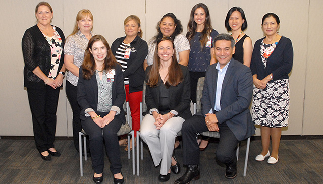
The Meyer Center for Developmental Pediatrics at Texas Children’s Hospital, Baylor College of Medicine and the Spina Bifida Association recently hosted the Houston Spina Bifida Education Day for patients, families and caregivers. Held at Houston Methodist Hospital, the event showcased newly established, evidence-based national guidelines for the care of individuals living with spina bifida. This is the first time the guidelines, written in part by Texas Children’s and Baylor clinicians and researchers, were shared with such a broad audience.

More than 100 people from across Texas and other states as far as Florida and Indiana attended the event. In addition to unveiling the new guidelines, experts from Texas Children’s presented talks covering a range of health issues faced by individuals living with spina bifida from infancy to adulthood. There also were presentations on related topics such as care coordination, emotional wellness, skin, and bowel-management tips specifically geared toward this group of families.

“Usually, clinical care guidelines are widely shared and disseminated among medical practitioners, not so much the lay public, so this conference not only provided professional guidance, but also much needed peer-to-peer advice,” said Dr. Ellen Fremion, a physician in the Spina Bifida Program at Texas Children’s and one of the organizers of the event. “Moreover, the heartfelt appreciation and deep emotional reactions from the families who attended this conference exemplifies how crucial it is to empower patients, families and caregivers with this kind of information, so they are better equipped to care for themselves and their family members.”

Spina bifida (meaning “split bone”) is the most common permanently disabling birth defect seen among newborns in the United States. It is a type of neural tube defect that occurs when a baby’s neural tube fails to develop or close properly. The symptoms of this condition range from mild to severe, depending on where the spinal cord is affected. Myelomeningocele is the most severe form of spina bifida in which parts of the spinal cord and nerves come through the open part of the spine. This leads to several related problems such as loss of feeling in areas below the opening, weakness or paralysis of the feet or legs, problems with bladder and bowel control. Some affected individuals have additional neurological complications, including a buildup of excess fluid around the brain (hydrocephalus) and diverse cognitive challenges. Multiple environmental (such as folic acid deficiency) and genetic factors are thought to cause this complex condition, although the exact causes are still unclear.

The Meyer Center’s Spina Bifida Program at Texas Children’s is a multidisciplinary program that includes several specialty services: neurosurgery, developmental pediatrics, urology, orthopedics, and physical medicine and rehabilitation. Experts from these specialties offer prenatal evaluations and follow patients from in utero to adulthood.

In 2011, Texas Children’s was among the first centers to perform open fetal surgery to treat spina bifida. Using this procedure, the defect in the fetal spine is accessed and repaired through an incision across the mother’s uterus (womb), and has since been the standard of care for spina bifida. In 2014, Texas Children’s Fetal Center pioneered a novel minimally-invasive fetoscopic procedure, in which the spinal defect is repaired through tiny incisions in the uterus using a small camera. This offers the same improved outcomes as the open fetal surgery but comes with additional benefits and reduced health risks.

“Despite recent advances of in utero repair procedures, surgery is not suitable for all patients and cannot be considered a cure,” said Dr. Jonathan Castillo, clinical director of the Spina Bifida Program at Texas Children’s. “Additionally, these surgeries may not reverse all the function or correct all related impairments. Also, these procedures are specific to myelomeningocele and may not help patients with other forms of spina bifida. Therefore, a deeper understanding of the etiology and biology of this condition is crucial to develop better treatment approaches in spina bifida care.”

In addition to providing cutting-edge clinical services, physicians and researchers at Texas Children’s Spina Bifida Program also are engaged in comprehensive, multidisciplinary research initiatives to find newer and safer medical and surgical interventions that can improve the quality of life for these individuals.

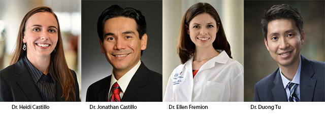
In 2014, a multidisciplinary team of experts at Texas Children’s Spina Bifida Program led by Drs. Heidi Castillo, Jonathan Castillo and Duong Tu, received a federal grant from the U.S. Centers for Disease Control and Prevention (CDC) to develop national standards in spina bifida care at the population level. Texas Children’s was the only funded hospital in Texas to be included in this effort, and, along with Baylor, was among the first institutions in the state to participate in the CDC’s National Spina Bifida Registry that collects medical data from this population to improve outcomes.

“Receiving that grant from the CDC and joining the registry gave us an amazing opportunity to participate in a nation-wide effort to develop better outcomes, interventions and standards of care for individuals with spina bifida,” Castillo said. “Since we are one of the major referral sites for spina bifida in the state of Texas, our team provides the entire spectrum of care to pediatric patients – from prenatal diagnosis, surgical interventions and postnatal care until adulthood – for myriad health issues related to spina bifida. This gives our team access to a wide demographic of participants and allows us to conduct unique in-depth studies and comparisons of the outcome measures of current interventions.”

“These studies have led to important findings that are reflected in the recent clinical care guidelines, and have revealed consistent disparities in the outcomes based on patient’s socioeconomic conditions and ethnicity, a novel observation that is currently under further investigation,” Castillo said. “Our team is excited that recently the CDC granted an extension of this grant for five more years, so we can continue to be at the forefront of comprehensive clinical care and research initiatives that will improve the lives of individuals living with spina bifida.”

September 9, 2014

91014NTD640

Physicians at the Fetal Center completed their first fetoscopic procedure to repair spina bifida in-utero – an innovative approach that was developed by Dr. Michael Belfort, obstetrician and gynecologist-in-chief at Texas Children’s Pavilion for Women and Dr. William Whitehead, pediatric neurosurgeon at Texas Children’s.

“Our hope is that these types of innovations will lead us to a new era of fetal medicine and surgery,” Belfort said.

Myelomeningocele, or open neural tube defect (NTD), is a form of spina bifida and occurs in 3.4 out of every 10,000 live births in the U.S. and is the most common permanently disabling birth defect for which there is no known cure. Myelomeningocele is a developmental defect in which the spine is improperly formed and the spinal cord is open to, and fused with, the skin. It is usually associated with hydrocephalus, or the buildup of cerebrospinal fluid in the brain, which requires surgical treatment to drain the fluid via an implanted device called a shunt. Originally, closure of the defect occurred after the birth of the baby, which was associated with an 80-90 percent chance that a shunt would be required for life.

But after a breakthrough NICHD-funded study entitled the Management of Myelomeningocele Study (MOMS) demonstrated a significant decrease in the risk of hydrocephalus for select patients undergoing fetal closure of the spine, as well as possible improvement in lower extremity function, compared to patients who underwent standard closure after birth, the Fetal Center adopted this as a treatment option and began performing open fetal surgery to treat spina bifida. To date, we still have one of only a handful of centers in the country that is able to perform this complex in-utero repair. Texas Children’s Fetal Center has completed 23 of these surgeries since 2011, with excellent results.

This technique, the standard of care in the US, involves a uterine incision and can cause maternal complications. With this in mind, the team at the Fetal Center wanted to focus on reducing the risks to the mother and countering the risks of preterm delivery. Working in conjunction with Dr. Jose Luis Peiro and Dr. Elena Carreras of Vall D’Hebron Hospital in Barcelona, Spain, they developed a fetoscopic approach to the repair.

The surgery was over three years in the making and features an in-utero, single layer, sutured repair through only two, four millimeter incisions in the uterus (rather than the 5-6cm opening that is required for an open procedure). In order to practice and perfect performing the closure using a minimally invasive fetoscopic approach, the team built a simulator using a child’s kickball that replicated the mother’s uterus. Inside the kickball, a doll acted as the fetus including a spina bifida defect on the doll’s back. Drs. Belfort and Whitehead practiced closing the defect using a team approach in which they both carry out specific parts of the surgery in a coordinated fashion. The two surgeons performed more than 30 simulated procedures including two full simulations, gowned and gloved, under actual OR conditions with a full support team.

“We have a magnificent team of specialists from a number of departments working together in the best interests of our fetal and neonatal patients. I am incredibly proud to be a member of this outstanding team and to be able to play a role in this mission,” Belfort said.

With a multi-disciplinary structured program in place, clinical planning and training and full Institutional Review Board approval for this experimental procedure, the team performed the first in-utero spin bifida closure on their first patient on July 30, 2014. The procedure went as planned and so far mom and baby are doing well. Doctors are optimistic and are waiting to access the outcome of the repair once the baby has been delivered.

“It is important to note that this mother has not been exposed to a significant uterine incision and we are much less concerned about her risk of a ruptured uterus during this pregnancy than we are after an open repair,” Belfort said. “We look forward to an uneventful delivery and a healthy baby.”

In addition to an expert operating room team and nursing staff, a multidisciplinary team of specialists led this first surgery at Texas Children’s Fetal Center, including Dr. Michael Belfort, Dr. William Whitehead, Dr. Alireza Shamshirsaz, Dr. Oluyinka O. Olutoye, Dr. Olutoyin Olutoye, Dr. David Mann, and Dr. Rodrigo Ruano.

“With the ever-advancing technology and imaging capabilities and dedicated surgeons, I am excited to see what the future holds when it comes to repairing anomalies fetoscopically,” Belfort said. “I am so impressed by what can be achieved with our exceptional team.”

April 1, 2014

4214Tspinabifida640

On February 23, 2012, the Fetal Center announced the birth of baby Charlotte, the first baby at Texas Children’s to receive in-utero surgery to treat spina bifida. Now, a little more than two years later, the center is celebrating another milestone with 20 cases completed.

After years of treating spina bifida by a neurosurgical closure of the spine defect during the first days of life, research breakthroughs prompted the Texas Children’s Fetal Center team to begin performing in-utero surgery on patients with a prenatal diagnosis of spina bifida.

Myelomeningocele, also known as spina bifida or open neural tube defect (NTD), is a developmental defect in which the spine is improperly formed and the spinal cord is open to and fused with the skin; it is usually associated with hydrocephalus, which requires surgical treatment to drain the fluid via a shunt. Myelomeningocele occurs in 3.4 out of every 10,000 live births in the U.S. and is the most common permanently disabling birth defect for which there is no known cure.

Advancements in fetal imaging and early prenatal diagnosis allow the team to gain access to a fetus while still inside a mother’s womb. Texas Children’s Fetal Center has developed extensive screening and diagnostic algorithms for pregnancies with fetal spina bifida. These algorithms determine which patients are appropriate candidates for the in-utero surgery.

The surgery is a huge, multi-disciplinary effort, bringing together a team of specialized experts, who all play a vital role in the medical treatment of both mom and baby. The team includes maternal-fetal specialists, led by Dr. Michael Belfort, fetal surgeons, led by Dr. Darrell Cass and Dr. Oluyinka Olutoye, and a neurosurgery team, led by Dr. William Whitehead. Once the fetus is exposed, Dr. Whitehead and his team surgically close the spinal defect before fetal surgeons return the baby to the womb. This procedure reduces the risk of hydrocephalus and may improve motor function in select patients.

A NICHD-funded study entitled the Management of Myelomeningocele Study (MOMS) published in the New England Journal of Medicine demonstrated a significant decrease in the risk of hydrocephalus for select patients undergoing fetal closure of the spine, as well as possible improvement in lower extremity function, compared to patients who underwent standard closure after birth. The MOMS trial is the second fetal intervention that has proved beneficial through a multi-center randomized clinical trial. The first was the Euro FETUS trial for laser ablation in the treatment of twin-to-twin transfusion syndrome (TTTS).

“Breakthrough studies like the MOMS trial are exciting and reaffirm our commitment to advancing fetal medicine and giving babies with complications and anomalies the healthiest possible start to life,” said Dr. Darrell Cass, co-director of Texas Children’s Fetal Center. “Our program has grown exponentially since we completed our first NTD repair in 2012, and we remain cautious, but it seems as though our center’s results are exceeding even those of the trial.”

The final piece to this puzzle is Dr. Kathryn Ostermaier, clinic chief of the Spina Bifida Clinic. It is under her guidance that the hospital provides long term care to surgical patients. The hospital’s Spina Bifida Clinic is a specialized program that serves only those patients with spina bifida, and includes several specialty services: orthopedics, neurosurgery, pediatrics, physical medicine and rehabilitation and urology.

The team that cares for NTD patients before birth, after birth and into childhood includes physicians from maternal fetal medicine, pediatric surgery, neurosurgery, anesthesiology, neonatology, pediatric radiology, cardiology, orthopedics, urology, physical therapy, and a highly dedicated group of specialized nurses, ultrasound technologists and genetic counselors.

“The confirmation that fetal surgery may decrease the physical challenges some of these babies face is not only a ray of hope for families, it is also a significant achievement for fetal medicine,” said Cass.