December 18, 2023

Magnetic resonance imaging (MRI) exams are an extremely valuable tool used in diagnosing patients. The exam length and unusual environment can be a cause of anxiety. Historically, pediatric patients needed general anesthesia to complete their exam, but through the MR-I Got This program, patients can use MRI mock scanners to help ease anxiety.

The mock scanners look and sound like a real scanner, but on a smaller scale. It has a bed that patients can lie on and travel into a tunnel similar to a real scanner. An MRI scanner makes loud noises while taking pictures, and the mock scanner simulates those noises preparing a child for what to expect. Allowing a child to practice their scan before they enter the real scanner helps them better understand their role and become more comfortable with something unfamiliar to them.

Since the launch in March 2022, MR-I Got This has had nearly 800 patients try the mock MRI scanner at both Medical Center and West Campus combined. Of those patients, 90% have been successful in completing their MRI without any medication. Additionally, the age range of patients completing this has been very wide with the youngest successful MR-I Got This patient at 2 ½ years old fully completing the MRI scan with the help of our amazing team.

“One of the biggest challenges we face with MRIs is getting the patient comfortable with the procedure to yield the best results,” said Dr. Nilesh Desai, Chief of Neuroradiology. “The mock scanner technology and the knowledge and patience of our team members has made all the difference in the progress we’ve made.”

This program is open to all patients free of charge and is available at both Medical Center and West Campus Radiology. Providers or patients can call to schedule the mock scanner and their non-sedate MRI to follow at the following locations:

  • Medical Center: 832-826-1334
  • West Campus: 832-227-2039

Radiologist Dr. Brandon Fuqua added, “It’s not a requirement to use the mock scanner, but the patients who benefit most are those who’ve never had an MRI before or experience anxiety. However, it’s open to anyone who feels it would benefit them.”

The mock scanner team consists of MRI Coordinators, Child Life, MRI Technologists and nurses all of whom help the patient through the process and answer questions they may have.

Click here to watch a video for more information or email MRIGotThis@texaschildrens.org.


November 18, 2021

From the vibrant walls and new furniture to the artwork adorning the hallways and ceilings (so patients can see it when they are lying on a gurney), the new Radiology floor is an impressive sight. But the move to the 18th floor of West Tower is so much more than a cosmetic improvement. The project was part of the Care First initiative and was delayed due mainly to the COVID-19 pandemic. The team has eagerly awaited the chance for patients to take advantage of the new space and everything it has to offer.

“We’re very excited to offer our patients an elevated level of service with our new facilities and technology,” said Dr. Thierry Huisman, Radiologist-in-Chief. “We have procedures and treatments no one else in the medical center can provide, and we will continue to stay on the cutting edge when it comes to providing the best care for our patients.”

The new magnetic resonance guided high intensity focused ultrasound (MR-HIFU) is the only one of its kind at both Texas Children’s Hospital and in The Texas Medical Center. It provides a much less invasive solution to remove tumors without radiation or surgery. In adults, it’s been used to reduce and even eliminate tremors.

The department will also house a Spectral CT scanner to offer much less invasive, yet superior molecular imaging with much lower radiation exposure to the patient. Plus, there are multiple large screen monitors in each room making it easier for the radiologist and treating physician/surgeon to communicate and view results in real-time.

The Siemens Myabi suite utilizes a full-scale CT scanner capable of moving between either IR room via rails built into the floor.

In addition, with the new space the number of PACU beds available increased from six to 16, nearly tripling the amount of patients Radiology can prep and recover. The existing space on the first floor will be used to expand the ER in the future.

——————–
National Radiologic Technology week ran recently from Nov. 7-13 to commemorate the discovery of the X-ray and its contributions to the medical and health sciences. The discovery of the X-ray advanced medical science by helping the diagnosis of fractures, broken bones and other ailments impacting the skeleton. Thank you to all the radiologic technologists at Texas Children’s for your contributions and continued dedication to our mission.

November 11, 2021

Dr Ananth Annapragada, vice chief of Research in the Edward B Singleton Department of Radiology, professor of Radiology and Obstetrics & Gynecology and program director of Basic Science Research at Texas Children’s Hospital and Baylor College of Medicine, has been named as a fellow of the National Academy of Inventors.

Annapragada is one of 175 notable academic inventors to receive this honor. His election was announced at the end of 2020 and he was recently honored during the 10th Annual NAI Meeting in Tampa, Florida.

Among his many accolades, Annapragada is also a member of the Dan L. Duncan Comprehensive Cancer Center, a fellow of the American Institute of Medical and Biological Engineers and a standing member of the NIH Nanotechnology (NANO) Study Section. His research primarily focuses on the development of novel technologies to address driving biological and medical questions. The innovative technologies he and his team develop include imaging for various cancers and Alzheimer’s disease, 3D Bioprinting and computational biology such as artificial intelligence and natural language processing.

Annapragada holds several patents, including 15 in the United States and nearly 100 worldwide with the majority of them in next generation imaging technologies, CT vascular imaging and MR molecular imaging.

The NAI Fellows Program highlights academic inventors who have demonstrated a spirit of innovation in creating or facilitating outstanding inventions that have made a tangible impact on the quality of life, economic development, and the welfare of society. Election to NAI Fellow is the highest professional distinction accorded solely to academic inventors.

August 17, 2020

For patients like 5-year-old Legend Pruitt, a visit to the doctor’s office can be a scary place for a child, especially if they don’t know what to expect when they come in for a medical procedure. A few months ago, when Legend had his MRI at Texas Children’s, his mother was surprised by how well he did, and how calm he was during the scan.

“He didn’t move at all because he knew he had to stay still for the MRI,” said Legend’s mother Niayeshua Joseph. “When my son had MRI scans before, he would have to be placed under anesthesia, because he could not stop moving. He is now more comfortable and the noises from the MRI machine don’t bother him.”

In an innovative effort to decrease the need for anesthesia in our MRI patients, Texas Children’s Radiology team bought the organization’s first mock MRI scanner last year thanks to a generous donation from one of our patient families. The mock MRI scanner provides a realistic simulation of what patients can expect before the real exam.

“This innovation allows patients to practice their scan before they enter the real scanner so they can understand their role (to lie still) and are more comfortable with something that is unfamiliar to them,” said Texas Children’s Radiologist-in-Chief Dr. Thierry Huisman. “Mock MR scanner training and familiarity with the MR imaging procedure enhances patient experience, reduces patient and family anxiety, and optimizes patient cooperation which can improve image quality or may allow for imaging without the need for patient sedation or general anesthesia.”

The mock MRI scanner on the fifth floor of Texas Children’s Lester and Sue Smith Legacy Tower, looks and sounds like a real MRI scanner. It has a bed that patients can lie on and travel into a tunnel similar to a real MRI scanner. The MRI scanner makes loud noises while the pictures are being taken. The mock MRI scanner simulates those noises and prepares a child for what to expect before they are scanned using the real machine. One additional advantage is that the mock MRI scanner does not include the “big magnet” and allows patients and families to attend without limitations. Furthermore, the mock MRI scanner does not use costly time as the real MRI unit.

Patients can get prepped with the mock MRI scanner in two ways. If the radiology pre-call nurse identifies a patient through their screening, they can schedule an appointment with a child life specialist several days before their procedure. Patients can also test out the mock MRI scanner immediately before their MRI appointment.

After introducing the mock scanner in July 2019, Radiology has seen amazing conversion results. Since June of this year, 67 patients have used the mock MRI scanner and 39 MRI patients scheduled for general anesthesia were converted to non-sedate procedures.

“We are really excited about our initial success,” Dr. Huisman said. “We are grateful to have this resource available to our patient families, and incredibly grateful to the Cook family’s philanthropic donation to make this all possible.”

A generous gift to help others

Brady and Nancy Cook know first-hand the challenges patient families face when a young child has to undergo an MRI especially for the first time. When their daughter, Alyssa, was in kindergarten, she started having complex partial seizures and was later diagnosed with a brain tumor. The brain tumor was believed to be in the area of the brain where speech is generated but doctors didn’t know for sure unless they performed a functional MRI on her.

“With a functional MRI, the patient has to be awake in the MRI machine and respond to images that flash up on the screen inside the MRI without moving because it would distort the quality,” said Brady Cook. “For that reason, it’s typically not done on 5-year-olds, because they are unable to hold still for an hour and follow instructions like that.”

But everything changed when the couple came across a research study on the use of mock MRI scanners that taught kids how to be more comfortable around the MRI machine to the point where young children could endure it. Out of desperation, they got creative and built a mock scanner in their garage. They used a big refrigerator box, a dolly that you lay on to get underneath a car, a softball mask to simulate the cage that goes over the patient’s head, a school locker sized mirror, age appropriate flash cards, and downloadable sound effects mimicking an MRI machine.

“We practiced this on our daughter so she could get comfortable with the MRI machine,” said Cook. “Soon after, she was able to successfully complete a functional MRI without moving. Her MRI scans showed us that the tumor was nowhere near the area of her brain that generates speech, and therefore the tumor could be removed safely.”

Since the mock MRI scanner helped their daughter, the Cook family wanted Texas Children’s to have the capability of helping patients become more comfortable with the MRI machine. Through their generous donation to Texas Children’s, the family is grateful that the mock MRI scanner is making a positive impact on patients and families.

“As parents who have been through this before and the desperation we felt at the time, we would do anything to ensure patients are comfortable during this process leading up to surgery,” Nancy Cook said. “Now that we have a mock scanner here, we hope that it will continue to give parents and children added comfort and reassurance.”

May 20, 2020

After months of planning and renovating, the Department of Radiology is excited to announce that it has installed a brand new state-of-the art 3 Tesla MRI unit at Texas Children’s Hospital West Campus. The Siemens 3T unit is one of now two MRIs at West Campus.

The ultra-high magnetic field strength of 3 Tesla will allow for:

  • Higher resolution imaging with optimized contrast
  • Bio-matrix guided imaging for faster scanning
  • Improved and expanded functional MRI capabilities

In addition, the modern design and high-end technology has multiple additional advantages including:

  • Wide Bore to accommodate a better fit and enhanced accessibility to the patient
  • Virtual Cockpit – Siemens scanner access from other locations Texas Children’s locations.

“This is a very exciting step toward broadening the scope of highly specialized care we can offer our patients in the West Houston area,” said Radiologist-in-Chief Dr. Thierry A. Huisman. “One of our main goals in the Department of Radiology, and across the Texas Children’s system, is to provide the right care, at the right time at the right place. The addition of the Siemens 3T helps us do that more so than ever.”

The room where the new MRI unit is housed was designed specifically for children. Covered in an aquatic-themed mural, the soothing ambiance helps reduce anxiety and decrease the need for sedation.

“We also use MRI- safe video goggles designed specifically to assist patients in MRI with minimizing claustrophobia and anxiety,” Huisman said, adding that children can pick a movie to watch during their procedure. “These goggles create a 3D virtual entertainment environment while reducing MRI noise.”

Due to the use of this entertainment environment and child-friendly atmosphere, 70 percent of MRI exams are now performed without sedation.

The addition of the Siemens 3T MRI adds to what is already a comprehensive suite of radiology services offered at West Campus. What is now offered at the community hospital is:

  • MRI (1.5 T and 3T)
  • CT
  • Ultrasound
  • X-ray and Fluoroscopy
  • Interventional Radiology
  • Multiple subspecialty pediatric radiology expertise including Musculoskeletal Radiology, Neuroradiology, Cardiac Imaging and much more.

“I’m very excited about what this new addition will bring to the hospital, our medical staff, and most importantly, our patients,” Huisman said.

May 18, 2020

As Texas Children’s begins its fifth official week of phased reopening and redesign, the organization continues to focus on a careful, strategic plan that supports family-centered care for our patients and meets our organizational expectations around quality and safety.

What this means for our patients and families is that services will open in phases, not all at once. For employees, a phased reopening means some of us will resume a full work schedule either at home or at one of our Texas Children’s facilities sooner than others.

“Patience and prudence are key,” said Chief Information and Innovation Officer Myra Davis. “We want to ensure everything we do operationally is safe and volume-driven. This means that just as we flexed down to demand, we will also flex back up to demand, where appropriate. The next few weeks and months ahead will be a thoughtful balance between the two and will help pave the way toward a successful future.”

Davis, along with Surgeon-in-Chief Dr. Larry Hollier and Executive Vice President Dan DiPrisco, are leading the Phased Reopen and Redesign Command for Texas Children’s.

Signs of success

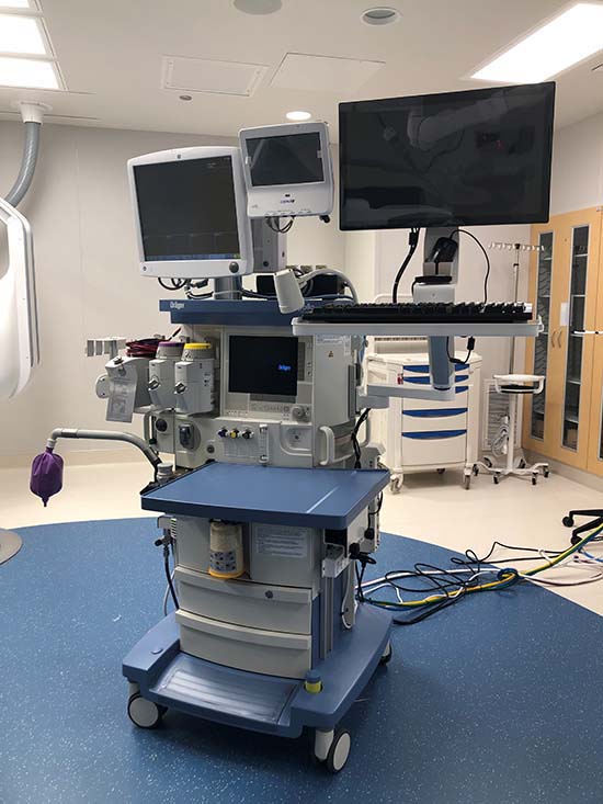
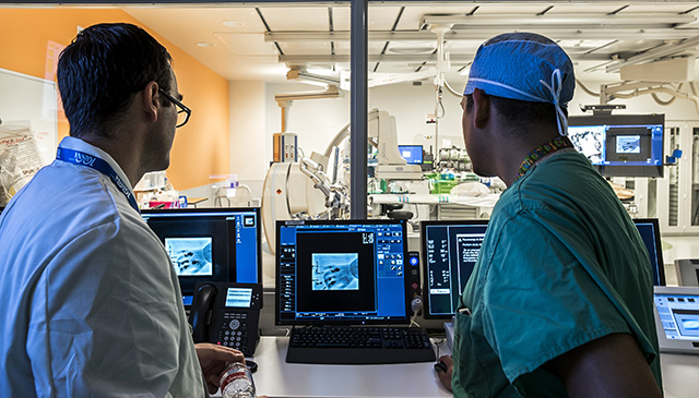
Two areas of our system that have seen early success during the phased reopening and redesign are Surgery and Radiology. Since reopening in late April, the Department of Surgery, Anesthesiology and Perioperative Services have opened schedules in operating rooms across the system to nearly 50 percent capacity. To date, 700-plus cases have been completed at our community locations and more than 1,000 pediatric and women’s cases have been conducted at the Texas Medical Center Campus.

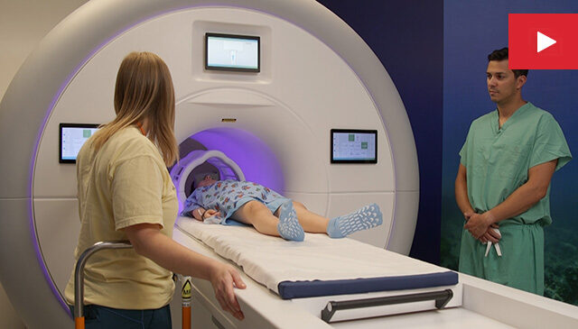
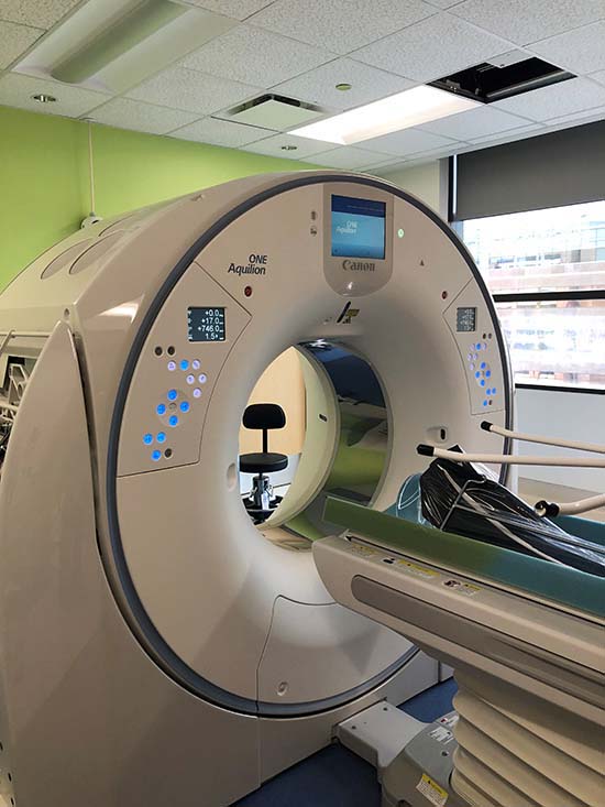
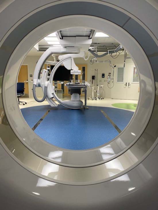
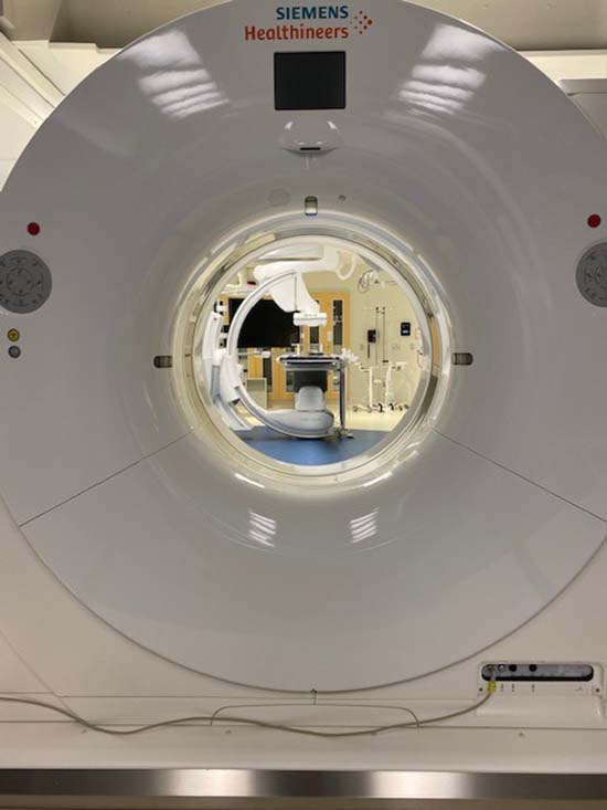
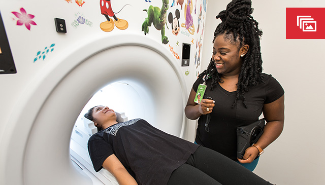
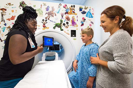
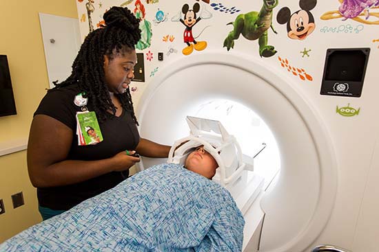
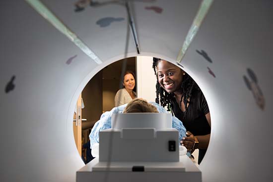
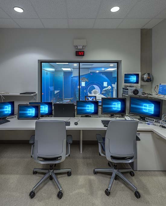
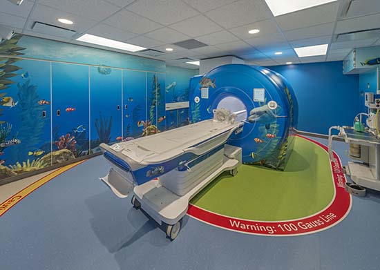
Two weeks ago, Radiology began its phased reopening and redesign, aiming for 50 percent of pre-COVID-19 imaging volume that week. The service line exceeded that goal, completing more than 640 studies per day. Pre-COVID-19, daily activity across the system was about 1,100 studies per day. During COVID-19, daily activity dropped to 350-400 studies per day. As a result of the steady uptick in patient volume, Radiology has completely reopened all of its appointments.

“We are encouraged by the quick response from the community to our thoughtful and agile reopening and redesign plans,” said Radiologist-in-Chief Dr. Thierry Huisman. “We are committed to continuing to serve our patients and families while keeping them and our staff safe and healthy during this pandemic.”

Hollier said his department plans to thoughtfully open additional ORs to match demand, all while maintaining social distancing protocols and other rigorous safety measures.

“Our primary goal has been and always will be to keep our patients and families safe and healthy,” he said.

Safety first

Safety measures and protocols have been put into place to protect our patients and families during their entire experience with Texas Children’s. This experience begins before they walk into one of our facilities and doesn’t end until long after they leave.

Before anybody comes into our facilities, they’re screened, to the best of our ability to make sure they are safe and healthy. This means all doctors, nurses, staff, patients and family members. Once they’re inside, everyone is required to wear a mask and adhere to social distancing.

“We want to respect these rules because we feel they protect patients from contracting the virus,” Hollier said. “We also feel it’s very important to test patients who are undergoing surgery and other procedures for COVID-19.”

No more than 48 hours before a surgical procedure, patients will receive a COVID-19 test, typically at one of our drive-through facilities for convenience. If the test is negative, the surgical procedure will proceed. If a patient tests positive for the coronavirus, the procedure will be delayed and retesting will occur to ensure the patient is negative before they undergo surgical or any other procedure that deems prior testing.

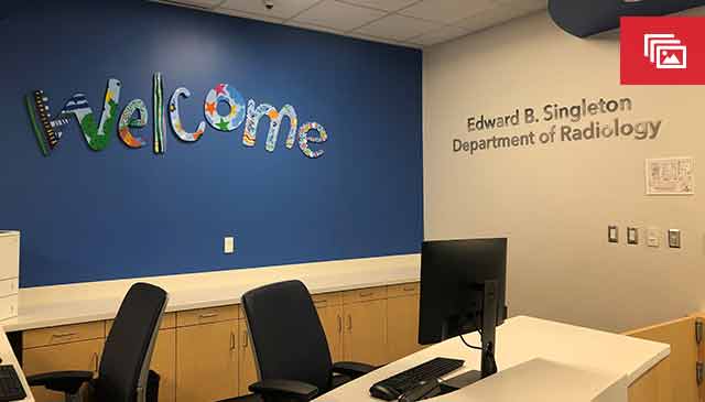
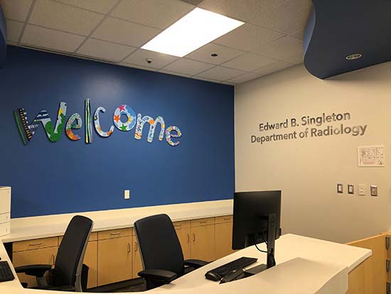
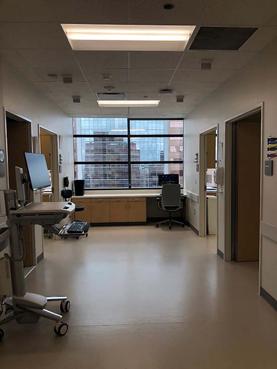
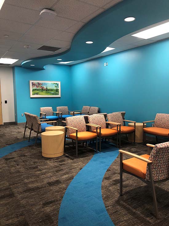
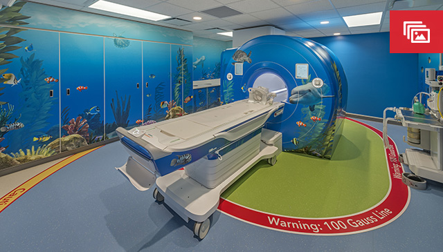
Radiology implemented a new process that allows patients to wait in their vehicle and call from the parking lot/garage upon arrival at our community locations, rather than entering the building and having to sit in the waiting area. Radiology services at Texas Children’s Hospital The Woodlands went live with pre check-in for patients who have a MyChart account. After a brief trial period, Radiology will implement this at West Campus and at our Medical Center Campus as well.

A mom who had rescheduled her daughter’s MRI due to safety concerns related to COVID-19, said she was relieved to learn about all of the safety measures and protocols Texas Children’s has in place to protect patients and families.

“Everything was easy and very smooth,” the mom said. “I appreciated calling from the garage, and I felt very safe.”

To learn more about additional precautions Texas Children’s is taking to protect our patients, families and staff click here.

Moving forward

This week, we launched plans for the reopening of our Ambulatory services and are working to ensure our facilities can accommodate increased activity while maintaining infection control guidelines.

Ramping our services back up at Texas Children’s is a welcoming indicator. It’s exciting, but this is merely the start for us. We are reopening thoughtfully and in phases, based on where we have the most demand for our services.

It will take much more time and careful planning to reopen completely. But moving strategically is what will restore us for the long-term, and what will ultimately ensure our organization’s future success and sturdiness.

“Thank you for all you are doing,” DiPrisco said. “The care you are providing for our patients and their families through this evolving situation is outstanding and proves that our challenges of the past few months have made us even better equipped for what’s ahead.”

January 6, 2020

Aurora Ayala, an administrative supervisor for Radiology, recently received the Edward B. Singleton Award for Administration. This prestigious award acknowledges individuals within Radiology whose outstanding leadership and/or contribution to the mission of the department has a significant and positive impact on advancing patient care, education, research or Texas Children’s core values.

Ayala began working with the late Dr. Singleton at St. Luke’s in 1984, who this award is named after. In 1991 she was hired as the supervisor of the Business Office at Texas Children’s when the physician and administrative offices were still in the same suite. When Singleton was chief, he would start his day by asking for two individuals, with one of them being Ayala. Her responsibilities included overseeing the scheduling, transcription and front desk teams. Since then, Ayala has helped the department grow into the success that it is today.

Ayala inspires others with her positive attitude and willingness to help everyone. She organizes department-wide events that boost employee morale and coordinates the day to day activities of the leaders she works with. She handles conflicts with patience and professionalism and ensures tasks are completed at the highest standard.

The Edward B. Singleton award was named in memory of Dr. Singleton, an early pioneer in radiology and long-time physician at Texas Children’s.