October 21, 2019

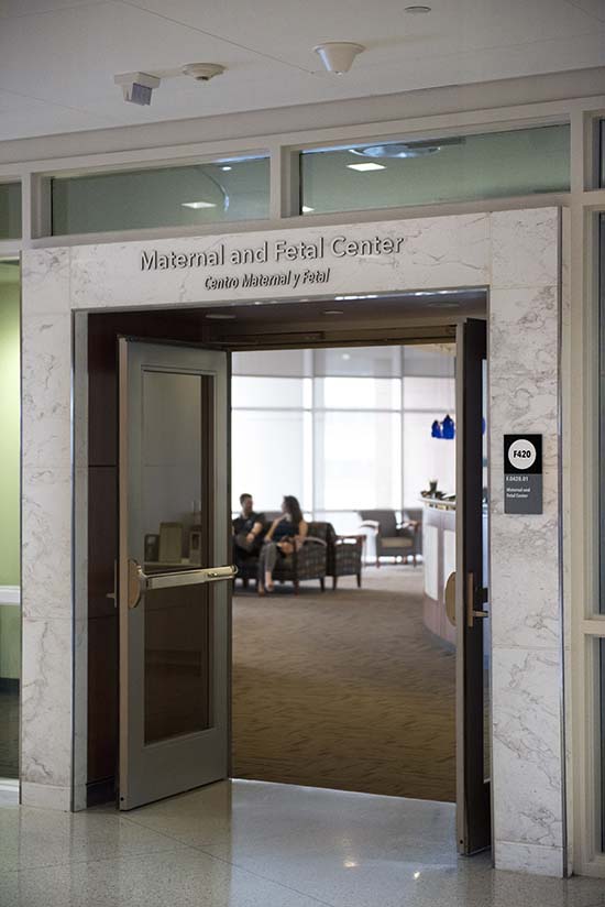
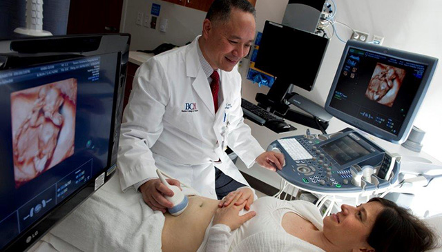
Due to a high demand for our expert maternal fetal medicine services and our goal to keep access open to all patients who need our care, Texas Children’s Pavilion for Women has expanded its Maternal Fetal Center.

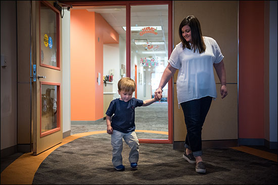
Located on the fourth floor of the Pavilion for Women, the center serves high-risk obstetrical patients and also bridges access to our pediatric subspecialists for our youngest patients through our Fetal Center program.

“The unit is designed so that the majority of services and partners needed to participate in a pregnancy come to us,” said Assistant Director of Ambulatory Clinical Practices for Women’s Services Aimee Jackson. “We pride ourselves on being a one-stop shop for our patients and families, providing timely, comprehensive, coordinated care in one place.”

Since opening its doors in 2012, the center has seen an 11 percent increase in patient visits. Last year, the center saw 36,000 patient visits and is expected to see 40,000 this year.

To continue to provide this growing patient population, construction on the Maternal Fetal Center began in September and was completed seven months ago at the end of April. The expansion added the following:

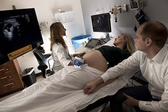
  • Three exam rooms bringing the total number of exam rooms in the center to nine. One of the exam rooms was built specifically for fetal intervention total care patients. These patients now can have their ultrasound, clinic visit and antenatal testing visit all in one location at the center. Historically, these patients had to schedule several different appointments in several different places to get these services.
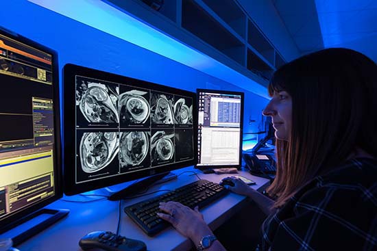
  • Two ultrasound rooms, bringing the total number to 11. One of the new can be used for clinical imaging as well as imaging research and educational training.
  • A second conference room equipped with advanced imaging display capabilities. The room will be used for team and family meetings as well as patient classes.
  • A large multi-person office space to accommodate the center’s expanded nursing and business support teams.
  • In addition, the center’s two existing reading rooms for radiology and cardiology were renovated, expanded and optimized.

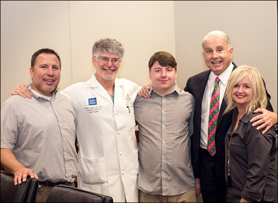
“We have needed this for a long time,” said Dr. Manisha Gandhi, the center’s medical director. “Most of our patients hoped they would never need our services, therefore we want to make their stay with us as comfortable as possible. This expansion will help us do that at an even higher level than we are now.”

Dr. Wesley Lee, chief of women and fetal imaging, agreed and said the center’s care team is committed to treating, respecting, and embracing patients with a spirit of hope and perseverance.

“Having the right space to carry out this type of care is key,” Lee said. “We are thrilled to have this extra space and to be able to use it to better serve our patients.”

Texas Children’s Pavilion for Women also is expanding its maternal fetal services at its community locations in the Greater Houston and beyond. Here are some of the latest developments:

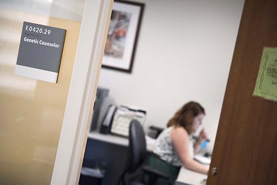
Baytown: In April, the Baytown Maternal Fetal Medicine Clinic moved to a larger space at Houston Methodist Baytown Hospital. The clinic – which offers maternal fetal consulting and ultrasound, as well as nutrition, genetic and psychiatric counseling – opened in 2016 for just one day a week. The number of patients coming to the clinic steadily increased, prompting leadership in 2018 to keep the clinic’s doors open five days a week. Today, the clinic’s team sees 10 to 15 patients a day. The majority of those patients, 70 percent, are seen by clinicians via telemedicine. The remaining 30 percent are seen by in-house medical staff.

Medical Center: In June, the Maternal Fetal Medicine Clinic in Houston Methodist Hospital’s Smith Tower in the Medical Center expanded from two ultrasound rooms to three and one non-stress test chair to two. As a result, the clinic can see more patients for Maternal Fetal Medicine services as well as nutrition and genetic counseling.

Lufkin: In November, a maternal fetal medicine clinic is slated to open at CHI St. Luke’s Health Memorial-Lufkin. Ultrasound and maternal fetal consults will be offered.

“Our aim with these community clinics is to provide excellent patient care closer to where some of our patients live,” said Jennifer Dalton, patient care manager for the Pavilion for Women Community Clinics. “We don’t want them to have to drive to the Medical Center unless they absolutely have to.”

Akachi Phillips, manager for the Pavilion for Women Community Clinics, said she knows a patient from Louisiana who drove weekly to the Medical Center for her visits during one of her pregnancies. During a subsequent pregnancy, she was able to have her ultrasounds in Baytown, which cut down her travel time and enhanced her overall experience.

“It means a lot to the patients to have a clinic nearby,” Phillips said.

For more information about the Maternal Fetal Center, click here.

August 26, 2019

Formerly conjoined twins Knatalye Hope and Adeline Faith Mata, who turned 5 on April 11, have started Pre-K 4 in Littlefield, Texas, marking a significant milestone four years after being separated in a more than 20-hour surgery at Texas Children’s Hospital.

Previously, the girls received various therapies, including physical, speech and occupational therapy and schooling in their home. Their mother, Elysse Mata, said the girls love their teachers and especially love recess. They’re also looking forward to taking the bus to school starting next week.

Following a complex pregnancy monitored by experts at Texas Children’s Fetal Center, the twins were safely delivered at Texas Children’s Pavilion for Women in 2014. After spending the first 10 months of their lives in the hospital’s neonatal intensive care unit, they underwent a successful separation surgery on Feb. 17, 2015. A team of more than 26 clinicians including 12 surgeons, six anesthesiologists and eight surgical nurses, among others, worked together to separate the girls who shared a chest wall, lungs, pericardial sac (the lining of the heart), diaphragm, liver, intestines, colon and pelvis.

During the complex surgery, the team worked for approximately 23 hours on Knatalye and 26 hours on Adeline, with the official separation occurring approximately 18 hours into the surgery. Among the surgical subspecialties involved were pediatric surgery, plastic surgery, cardiovascular surgery, urology, liver surgery, orthopedic surgery and pediatric gynecology.

Both girls are doing extremely well and continue to thrive at home where they live with their parents, Eric and Elysse, older brother, Azariah, and younger sister, Mia. The family is very excited to see the girls sharing this special milestone together.

August 20, 2019

Fetal growth restriction is a major public health concern that can lead to short-term complications for the newborn and possibly the development of health problems later in life. Researchers at Texas Children’s Hospital and Baylor College of Medicine were recently awarded $3.2 million by the National Institutes of Health to develop an improved way to assess umbilical venous blood flow using 3D and Doppler ultrasound techniques. They aim to improve the detection and monitoring of small fetuses.

Fetal growth restriction increases the risk of stillbirth, problems during the newborn period, and neonatal death. Affected fetuses also are predisposed to developmental delay as well as the occurrence of adult diseases such as obesity, type 2 diabetes mellitus, coronary artery disease, and stroke.

“Our research team will initially validate the accuracy and reproducibility of new 3D volume flow measurements and then develop corresponding reference ranges in normal pregnancies,” said Dr. Wesley Lee, co-director of Texas Children’s Fetal Center and professor of obstetrics and gynecology at Baylor. “Detailed observations of fetal growth, heart function, and circulatory changes will be made in over 1,000 small fetuses with estimated weights below the tenth percentile. The results will be correlated with pregnancy outcomes to identify prenatal predictors of clinical problems in newborns.”

According to Lee, identifying the most vulnerable, small fetuses may not only influence their neonatal course, but could also have lasting impact on long-term health consequences during adult life. Researchers hope to develop 3D umbilical venous flow as a reproducible circulatory measurement that is accurate and clinically applicable, even during early pregnancy.

The five-year investigation is a collaboration between Texas Children’s Hospital, Baylor College of Medicine, University of Michigan, Perinatology Research Branch of the Eunice Kennedy Shriver National Institute of Child Health and Human Development and GE Healthcare.

March 25, 2019

Almost seven years ago, surgeons with Texas Children’s Fetal Center performed their first in-utero neural tube defect repair surgery. Just a few months ago, some of those same surgeons reached a milestone when they completed their 100th neural tube defect repair.

Of the 100 total cases, more than half were performed fetoscopically, an experimental, minimally-invasive surgical approach pioneered at Texas Children’s in 2014 by Obstetrician and Gynecologist-in-Chief Dr. Michael Belfort and Pediatric Neurosurgeon Dr. William Whitehead.

Texas Children’s was the first center in the U.S. to correct neural tube defects, also known as spina bifida defects, fetoscopically and is among just a few centers in the country that offers fetoscopic repair of spina bifida. Texas Children’s continues to offer open fetal surgery for spina bifida, the standard of care since 2012, for patients who do not qualify for or opt not to undergo a fetoscopic repair.

“This milestone shows that we are a mature program, that we have done a lot of these cases, and that our results are equivalent if not better, than anybody else out there doing this,” Belfort said. “We have a level of experience now whereby we can assure patients and families that what we offer is the best in terms of quality and safety.”

Neural tube defect repair surgeries are performed on babies with Myelomeningocele, a developmental defect in which the spine is improperly formed and the spinal cord is open to and fused with the skin. The condition, also known as spina bifida or an open neural tube defect (NTD), occurs in 3.4 out of every 10,000 live births in the U.S. and is the most common permanently disabling birth defect for which there is no known cure.

NTDs are usually associated with motor impairment and hydrocephalus, or the buildup of cerebrospinal fluid in the brain, which requires surgical treatment to drain the fluid via an implanted device called a shunt. It’s common for children with spina bifida to have abnormal function of their bladder, bowels and legs. The goal of fetal surgical repair of spina bifida is to preserve leg function and reduce the need for a postnatal shunt.

The standard of care for spina bifida is neurosurgical closure of the defect in the first days of life, however, advances in fetal surgery and the landmark clinical trial, known as the Management of Myelomeningocele Study (MOMS Trial), proved that a fetal surgical repair leads to decreased rates of hydrocephalus and improved leg function compared to a postnatal repair.

Through their research and outcomes data, Belfort and his team have shown that performing the surgery fetoscopically yields the same outcome for the baby as the open repair, while being significantly less invasive for the mother.

“We are thrilled to offer this innovative and minimally-invasive surgical approach to our patients and their babies,” Belfort said. “We thank the mothers and families who put their trust in our team and have the courage to undergo an experimental procedure.”

First fetoscopic case

The first mother to undergo fetoscopic neural tube repair at Texas Children’s Fetal Center was Althea Canezaro. At 23 weeks pregnant, Althea learned during a routine ultrasound that her son, Grayson, had spina bifida. The Louisiana resident came to Texas Children’s shortly thereafter and met with Belfort and his team.

“After talking with them, it was like, OK, there’s something they can do for us, and if it didn’t work out everyone would be safe,” Althea recalled. “We knew that this was an opportunity to give him the best life that he could have. So, going forward, it was kind of like the ball was in their park. The weight was taken off our shoulders.”

A little more than four years later, Althea and Grayson are thriving. The blonde haired, blue-eyed boy never developed hydrocephalus and was born with full movement of his legs. He is hitting all of his developmental milestones and walks with and without the assistance of crutches.

“He’s a fighter,” Althea said. “You can’t tell him he can’t do anything. He has more determination than you ever want to believe.”

Reaching a milestone

Belfort and his team performed the 100th neural tube defect repair late last year and delivered the healthy little girl, Parker Kate, on March 5. Taylor Avera, the newborn’s mother, said when she and her husband found out their child had spina bifida they were scared to death, but that what they learned at Texas Children’s put them at ease.

“Neither one of us questioned that this was what we needed to do,” Taylor said. “We were excited that we were candidates for the surgery and that we were going to be able to give our baby the best outcome she could have.”

The experimental surgery Althea and Taylor underwent continues to be subject to rigorous oversight by the U.S. Food and Drug Administration, was developed by Texas Children’s Fetal Center in partnership with Texas Children’s division of neurosurgery. The teams practiced on a child’s kickball that replicated the mother’s uterus. Inside the kickball, a doll acted as the fetus. By hoisting the ball into the air hung by various levers and pulleys, the team simulated the movement in the womb and practiced closing the spinal cord.

“The multidisciplinary collaboration with the Fetal Center on this surgical effort is an extraordinary example of the innovative spirit at Texas Children’s Hospital,” Whitehead said. “From a medical standpoint, we believe by closing the defect both in-utero and fetoscopically, is a less invasive procedure for the mother, reduces her risk of preterm delivery and reduces the need for shunts.”

View photos of both Grayson and Parker Kate below.

Texas Children’s Fetal Center is among the nation’s leaders in providing high-risk maternal care and the diagnosis and treatment of abnormalities in unborn and newborn infants. For more information, visit women.texaschildrens.org/fetal.

August 27, 2018

Fetal surgeon and Ob-Gyn-in-chief Dr. Michael Belfort is often reunited with his patients, but the recent meeting he had with Sam Hancock was extra special. The teenager and his family made their way from Utah to Houston to meet Belfort, who saved Sam’s life before he was even born. The meeting was Sam’s 18th birthday wish and his parents, Alisa and Dennis, were happy to accommodate as Belfort’s name holds a high regard in their home.

“As a family, we are so grateful for Dr. Belfort,” Alisa said. “He deserves the credit for the skill and knowledge that gave Sam a chance at life.”

When Alisa was pregnant with Sam nearly two decades ago, an ultrasound showed severe swelling of her son’s neck. Doctors told her and Dennis their baby likely had a very serious condition and would not survive.

Later in her pregnancy, doctors noticed fluid building up in Sam’s chest cavity and told her there was no hope. That’s when the family found Belfort, who was practicing in Utah at the time and gave them the option of having fetal surgery. Belfort placed a shunt into Sam’s chest in hopes that fluid being produced would drain from his chest into the amniotic sac, relieving the pressure on his developing heart and lungs. The shunt worked for a short time, but then stopped. Belfort made a second attempt and it was successful in draining the fluid and taking the pressure off Sam’s heart and lungs.

Sam was delivered two months early as Alisa went into early labor because of extra fluid in her amniotic sac, a condition called polyhydramnios, which stretches the uterus making it extremely large. Despite several attempts to decrease the amount of fluid, labor could not be stopped.

Sam spent three months in the neonatal intensive care unit and underwent another surgery to place drains in his chest to relieve excess fluid. When he was discharged, Sam was on oxygen. At 2 years old, he was diagnosed with craniosynostosis, a condition where the skull bones are fused and cause a misshapen head. To correct the condition and to relieve pressure on his brain, Sam underwent yet another surgery.

Despite a rough start in life, Sam is a fighter. Though he battled some academic delays earlier in life, he worked hard to overcome his challenges and recently graduated with a 3.5 GPA and earned his Eagle Scout award. When asked what he wanted for his 18th birthday, he said he wanted to come to Houston to see Belfort, and to thank him for saving his life and never giving up on him.

“There are no words to express what I’m feeling right now,” Sam said after shaking Belfort’s hand and grinning ear to ear. “I am so happy to be here.”

Belfort said meeting back up with Sam and his family was a real treat and that he had never forgotten them.

“It was an unusual procedure at that time,” Belfort said of the operation he did on Sam more almost two decades ago. “I’m really glad it worked out. Sam is a delightful young man.”

During his visit to Houston, Belfort gave Sam and his family a tour of Texas Children’s Hospital and introduced them to Texas Children’s President and CEO Mark Wallace, who was so honored to meet Sam he personally sang Happy Birthday to the teenager.

After talking to Sam about his hopes and dreams, Wallace told him about his 10 leadership maxims and encouraged him to come up with his own personal definition of leadership.

“Now that you are 18, you are old enough to have your own definition of leadership,” Wallace said. “That definition should reflect you, your personality and your beliefs.”

Sam, who is looking forward to the next journey in his life and will soon be looking for a job, promised Wallace he would work on coming up with his personal definition of leadership and share it with him soon.

May 1, 2018

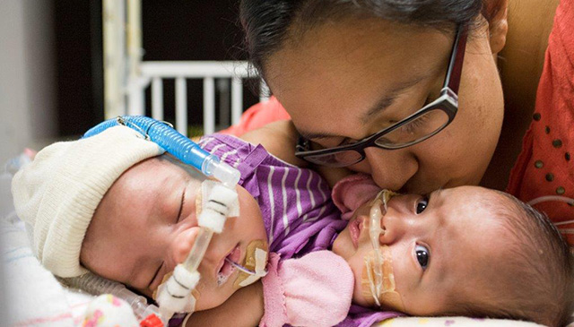
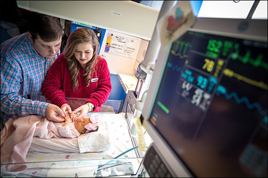
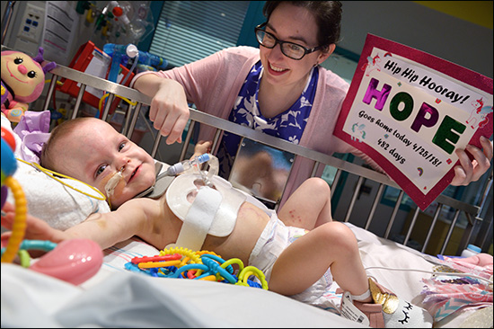
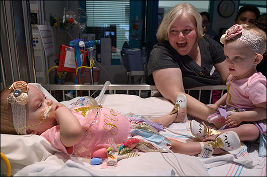
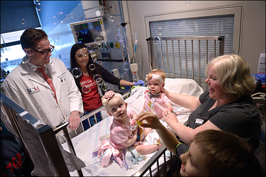
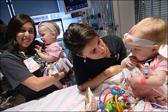
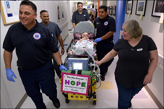
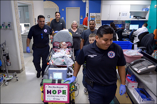
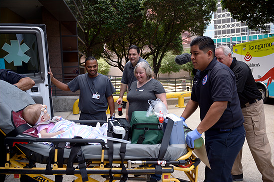
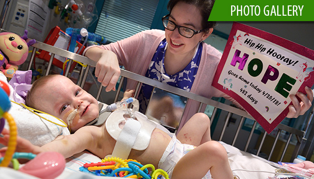
Hope Elizabeth Richards, one of the formerly conjoined twin girls separated at Texas Children’s earlier this year, was discharged April 25 after spending 482 days in the hospital. Hope joined her sister, Anna Grace, who was discharged on March 2.

The Richards family is looking forward to returning to their North Texas home soon. They are grateful for all of the support and prayers they received throughout their daughters’ journey.

“This is the moment it all feels real,” said Jill Richards. “We are so excited for Hope to join Anna and her brothers at home. Our family is eternally thankful for the doctors, nurses, child life specialists, physical therapists and many others at Texas Children’s who took incredible care of our precious girls.”

On January 13, Anna and Hope were successfully separated by a multidisciplinary team of nearly 75 surgeons, anesthesiologists, cardiologists and nurses from eight specialties performed the seven-hour procedure. In preparation for separation, on November 6, 2017, Anna and Hope underwent surgery to place tissue expanders in order to allow their skin to grow and stretch.

The girls were born on December 29, 2016 at Texas Children’s Pavilion for Women, weighing a combined 9 lbs. 12 oz. Delivered via Cesarean-section at 35 weeks and five days gestation, Anna and Hope were conjoined at their chest and abdomen, through the length of their torso and shared the chest wall, pericardial sac (the lining of the heart), diaphragm and liver. In addition, they had a large blood vessel connecting their hearts. They were welcomed by their parents, Jill and Michael, and older brothers Collin and Seth.

The Richards family, learned Jill was carrying conjoined twins during a routine ultrasound. The family was then referred to Texas Children’s Fetal Center, where they underwent extensive prenatal imaging, multidisciplinary consultation and development of plans to achieve a safe delivery and postnatal care. They temporarily relocated to Houston in order to deliver at Texas Children’s and to be close to the girls during their hospital stay. For the past year, Anna and Hope have been cared for by a team of specialists in the level IV and level II neonatal intensive care units (NICU).

April 3, 2018

On March 26, Dr. Wesley Lee, co-director of Texas Children’s Fetal Center, located within Texas Children’s Pavilion for Women, was presented with the Joseph H. Holmes Pioneer Award at the American Institute of Ultrasound in Medicine’s (AIUM) 2018 convention in New York. Each year, the award honors at least two current or retired AIUM members whose clinical or basic science work significantly contributed to the growth and development of medical ultrasound.

Among his many accomplishments, Lee has authored 148 peer-reviewed articles and 20 book chapters pertaining to maternal-fetal medicine, prenatal detection of congenital anomalies, 3-/4-dimensional fetal sonography and fetal magnetic resonance imaging.